 Explore
 Validate
 Learn
Learn Western blot
Antibody data
- Antibody Data
- Antigen structure
- References [1]
- Comments [0]
- Validations
- Western blot [4]
- Immunohistochemistry [1]
Submit
Validation data
Reference
Comment
Report error
- Product number
- HPA008188 - Provider product page

- Provider
- Atlas Antibodies
- Proper citation
- Atlas Antibodies Cat#HPA008188, RRID:AB_1855819
- Product name
- Anti-PSMA2
- Antibody type
- Polyclonal
- Reactivity
- Human, Mouse, Rat
- Host
- Rabbit
- Conjugate
- Unconjugated
- Antigen sequence
SGAYFAWKATAMGKNYVNGKTFLEKRYNEDLELED
AIHTAILTLKESFEGQMTEDNIEVGICNEAGFRRL
TPTEVKDYLAAIA- Isotype
- IgG
- Vial size
- 100 µl
- Storage
- Store at +4°C for short term storage. Long time storage is recommended at -20°C.
Submitted references Initial Quantitative Proteomic Map of 28 Mouse Tissues Using the SILAC Mouse
Geiger T, Velic A, Macek B, Lundberg E, Kampf C, Nagaraj N, Uhlen M, Cox J, Mann M
Molecular & Cellular Proteomics 2013 June;12(6):1709-1722
Molecular & Cellular Proteomics 2013 June;12(6):1709-1722
No comments: Submit comment
Supportive validation
- Submitted by
- Atlas Antibodies (provider)
- Enhanced method
- Genetic validation
- Main image

- Experimental details
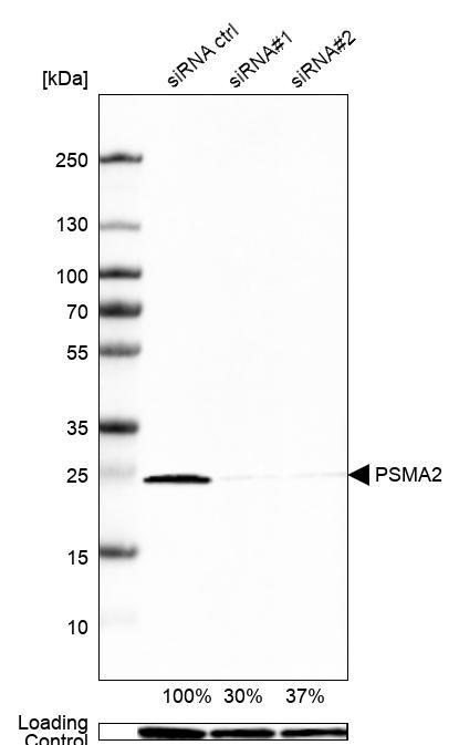
- Western blot analysis in U2OS cells transfected with control siRNA, target specific siRNA probe #1 and #2, using Anti-PSMA2 antibody. Remaining relative intensity is presented. Loading control: Anti-GAPDH.
- Submitted by
- Atlas Antibodies (provider)
- Main image

- Experimental details
- Lane 1: NIH-3T3 cell lysate (Mouse embryonic fibroblast cells)Lane 2: NBT-II cell lysate (Rat Wistar bladder tumour cells)

- Submitted by
- Atlas Antibodies (provider)
- Main image

- Experimental details
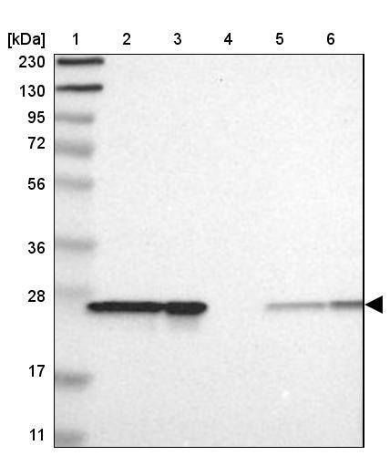
- Lane 1: Marker [kDa] 230, 130, 95, 72, 56, 36, 28, 17, 11Lane 2: Human cell line RT-4Lane 3: Human cell line U-251MG spLane 4: Human plasma (IgG/HSA depleted)Lane 5: Human liver tissueLane 6: Human tonsil tissue
- Submitted by
- Atlas Antibodies (provider)
- Main image

- Experimental details
- Western blot analysis in mouse cell line NIH-3T3 and rat cell line NBT-II.

Supportive validation
- Submitted by
- Atlas Antibodies (provider)
- Main image
- Experimental details
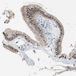
- Immunohistochemical staining of human gallbladder shows moderate cytoplasmic and nuclear positivity in glandular cells.
- Sample type
- HUMAN
