Explore
 Validate
 Learn
Learn Western blot
Antibody data
- Antibody Data
- Antigen structure
- References [1]
- Comments [0]
- Validations
- Western blot [2]
- Immunocytochemistry [1]
- Immunohistochemistry [5]
Submit
Validation data
Reference
Comment
Report error
- Product number
- PA5-52476 - Provider product page

- Provider
- Invitrogen Antibodies
- Product name
- SV2A Polyclonal Antibody
- Antibody type
- Polyclonal
- Antigen
- Recombinant full-length protein
- Description
- Immunogen sequence: SDGYYRGEGT QDEEEGGASS DATEGHDEDD EIYEGEYQGI PRAESGGKGE RMADGAPLAG VRGGLSDGEG PPGGRGEAQR RKEREELAQQ YEAILRECGH GRFQW
- Concentration
- 0.08 mg/mL
Submitted references Nicotinic acetylcholine receptors regulate clustering, fusion and acidification of the rat brain synaptic vesicles.
Trikash I, Kasatkina L, Lykhmus O, Skok M
Neurochemistry international 2020 Sep;138:104779
Neurochemistry international 2020 Sep;138:104779
No comments: Submit comment
Supportive validation
- Submitted by
- Invitrogen Antibodies (provider)
- Main image
- Experimental details
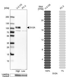
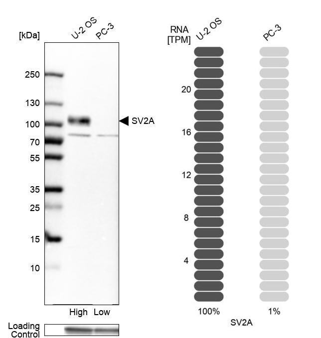
- Western blot analysis of SV2A in human cell lines U2OS and PC-3 using a SV2A Polyclonal Antibody (Product # PA5-52476). Corresponding SV2A RNA-seq data are presented for the same cell lines. Loading control: Anti-GAPDH.
- Submitted by
- Invitrogen Antibodies (provider)
- Main image

- Experimental details
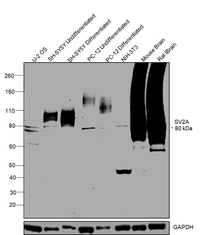
- Western blot was performed using Anti-SV2A Polyclonal Antibody (Product # PA5-52476) and a 80kDa band corresponding to SV2A was observed across all cell lines and tissue tested where there is heavy glycosylation in the brain tissues. Whole cell extracts (30 µg lysate) of U-2-OS (Lane 1), undifferentiated SH-SY5Y (Lane 2), differentiated SH-SY5Y (Lane 3), undifferentiated PC-12 (Lane 4), differentiated PC-12 (Lane 5), NIH-3T3 (Lane 6), Mouse brain (Lane 7) and Rat brain (Lane 8) were electrophoresed using NuPAGE® 4-12 % Bis-Tris gel (Product # NP0321BOX). Resolved proteins were then transferred onto a nitrocellulose membrane (Product # IB23001) by iBlot® 2 Dry Blotting System (Product # IB21001). The blot was probed with the primary antibody (0.4ug/ml) and detected by Goat anti-Rabbit IgG (H+L), Superclonal™ Recombinant Secondary Antibody, HRP conjugate (Product # A27036, 1:4000 dilution) using the iBright FL 1000 (Product # A32752). Chemiluminescent detection was performed using Novex® ECL Chemiluminescent Substrate Reagent Kit (Product # WP20005).
Supportive validation
- Submitted by
- Invitrogen Antibodies (provider)
- Main image
- Experimental details
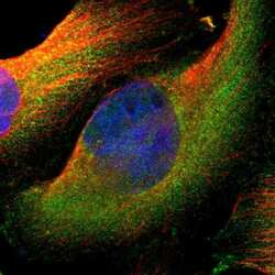
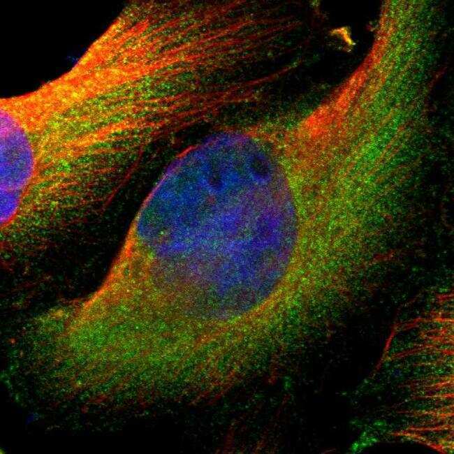
- Immunofluorescent staining of SV2A in human cell line U-251 MG shows positivity in cytoplasm. Samples were probed using a SV2A Polyclonal Antibody (Product # PA5-52476).
Supportive validation
- Submitted by
- Invitrogen Antibodies (provider)
- Main image
- Experimental details
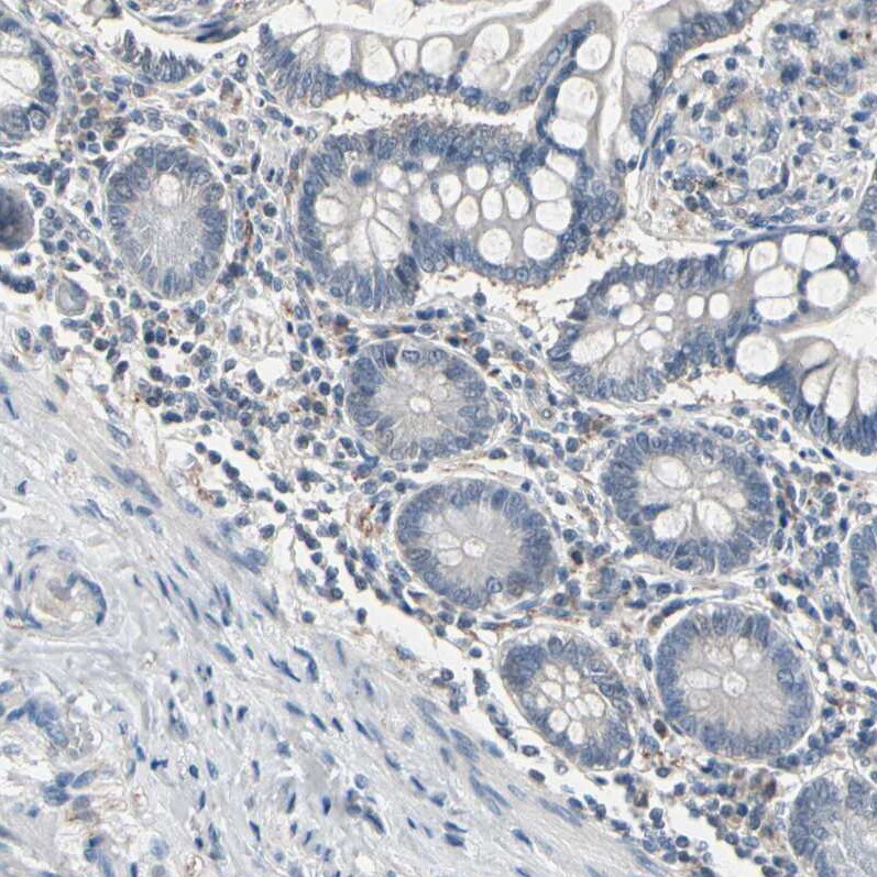
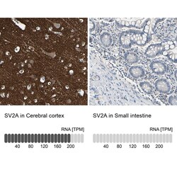
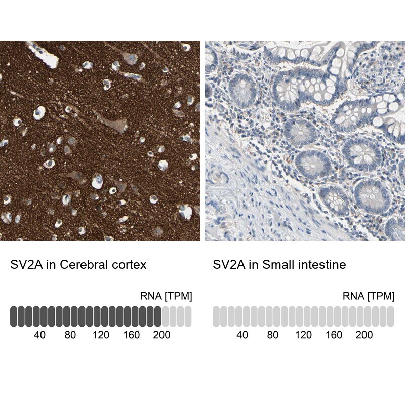
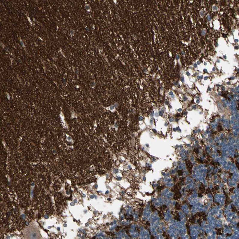
- Immunohistochemical staining of SV2A in human cerebral cortex and small intestine tissues using SV2A Polyclonal Antibody (Product # PA5-52476). Corresponding SV2A RNA-seq data are presented for the same tissues.
- Submitted by
- Invitrogen Antibodies (provider)
- Main image
- Experimental details
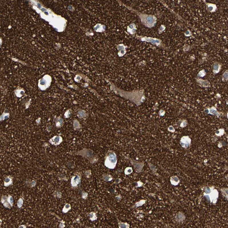
- Immunohistochemical staining of SV2A in human cerebral cortex using SV2A Polyclonal Antibody (Product # PA5-52476) shows strong cytoplasmic positivity in neuropil.
- Submitted by
- Invitrogen Antibodies (provider)
- Main image
- Experimental details
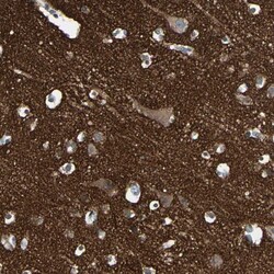
- Immunohistochemical staining of SV2A in human cerebellum using SV2A Polyclonal Antibody (Product # PA5-52476) shows strong cytoplasmic positivity in neuropil.
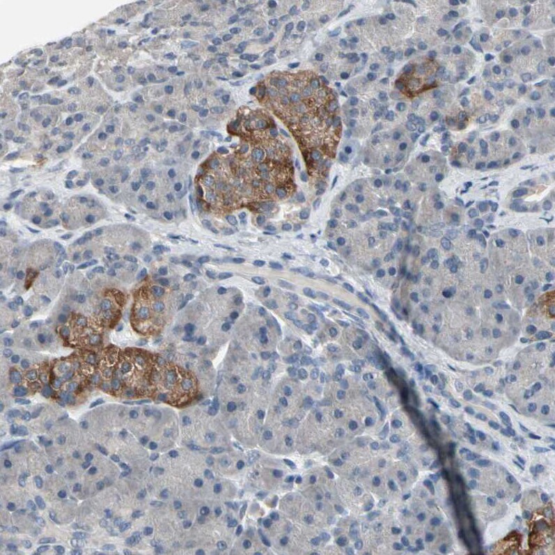
- Submitted by
- Invitrogen Antibodies (provider)
- Main image

- Experimental details
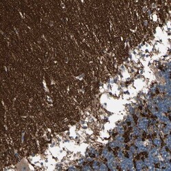
- Immunohistochemical staining of SV2A in human pancreas using SV2A Polyclonal Antibody (Product # PA5-52476) shows strong cytoplasmic positivity in islets of Langerhans.
- Submitted by
- Invitrogen Antibodies (provider)
- Main image

- Experimental details
- Immunohistochemical staining of SV2A in human small intestine using SV2A Polyclonal Antibody (Product # PA5-52476) shows no positivity in glandular cells.