 Explore
 Validate
 Learn
Learn Western blot
Antibody data
- Antibody Data
- Antigen structure
- References [0]
- Comments [0]
- Validations
- Western blot [3]
- Immunocytochemistry [1]
- Flow cytometry [1]
Submit
Validation data
Reference
Comment
Report error
- Product number
- PA5-47450 - Provider product page

- Provider
- Invitrogen Antibodies
- Product name
- IGSF8 Polyclonal Antibody
- Antibody type
- Polyclonal
- Antigen
- Recombinant full-length protein
- Description
- In direct ELISAs, approximately 15% cross-reactivity with recombinant human IGSF8 is observed and less than 1% cross-reactivity with recombinant mouse IGSF4 is observed. Reconstitute at 0.2 mg/mL in sterile PBS.
- Reactivity
- Human, Mouse, Rat
- Host
- Goat
- Isotype
- IgG
- Vial size
- 100 µg
- Concentration
- 0.2 mg/mL
- Storage
- -20° C, Avoid Freeze/Thaw Cycles
No comments: Submit comment
Supportive validation
- Submitted by
- Invitrogen Antibodies (provider)
- Main image
- Experimental details
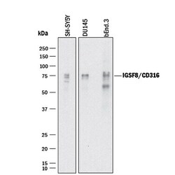
- Western blot analysis of IGSF8 in SH‚SY5Y human neuroblastoma cell line, DU145 human prostate carcinoma cell line, and bEnd.3 mouse endothelioma cell line. Samples were incubated in IGSF8 polyclonal antibody (Product # PA5-47450) using a dilution of 1 µg/mL followed by a HRP-conjugated Anti-Goat IgG secondary antibody. Specific bands were detected for IGSF8/CD316 at approximately 70-80 kDa (as indicated). This experiment was conducted under reducing conditions.

- Submitted by
- Invitrogen Antibodies (provider)
- Main image
- Experimental details
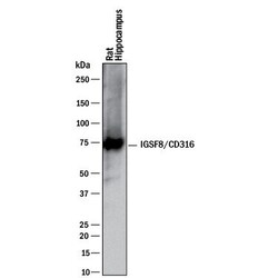
- Western blot analysis of IGSF8 in rat brain (hippocampus) tissue. Samples were incubated in IGSF8 polyclonal antibody (Product # PA5-47450) using a dilution of 1 µg/mL followed by a HRP-conjugated Anti-Goat IgG secondary antibody. A specific band was detected for IGSF8/CD316 at approximately 70 kDa (as indicated). This experiment was conducted under reducing conditions.

- Submitted by
- Invitrogen Antibodies (provider)
- Main image
- Experimental details
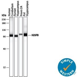
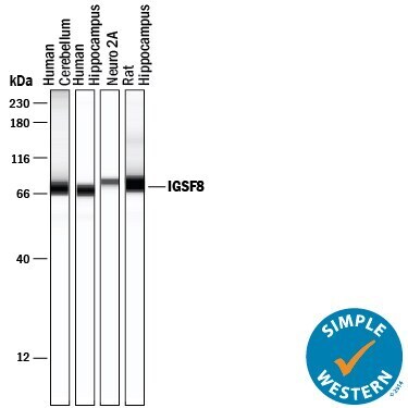
- Western blot analysis of IGSF8 in 0.2 mg/mL lysates of human cerebellum tissue, human hippocampus tissue, Neuro‚2A mouse neuroblastoma cell line, and rat hippocampus tissue. Samples were incubated in IGSF8 polyclonal antibody (Product # PA5-47450) using a dilution of 20 µg/mL followed by HRP-conjugated Anti-Goat IgG at a dilution of 1:50. A specific band was detected for IGSF8/CD316 at approximately 71-80 kDa (as indicated). This experiment was conducted under reducing conditions and using the 12-230 kDa separation system.
Supportive validation
- Submitted by
- Invitrogen Antibodies (provider)
- Main image
- Experimental details
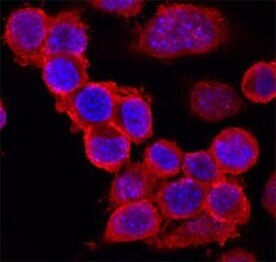
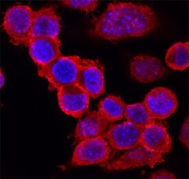
- Immunocytochemistry analysis of IGSF8 in immersion fixed Neuro‚2A mouse neuroblastoma cell line. Samples were incubated in IGSF8 polyclonal antibody (Product # PA5-47450) using a dilution of 10 µg/mL for 3 hours at room temperature followed by NorthernLights™ 557-conjugated Anti-Goat IgG Secondary Antibody (red) and counterstained with DAPI (blue). Specific staining was localized to cell surfaces.
Supportive validation
- Submitted by
- Invitrogen Antibodies (provider)
- Main image
- Experimental details
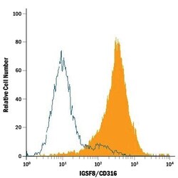
- Flow cytometry of IGSF8 in Neuro‚2A mouse neuroblastoma cell line. Samples were incubated in IGSF8 polyclonal antibody (Product # PA5-47450) or isotype control antibody followed by Phycoerythrin-conjugated Anti-Goat IgG Secondary Antibody.
