Explore
 Validate
 Learn
Learn Western blot
 Immunocytochemistry
Antibody data
- Antibody Data
- Antigen structure
- References [3]
- Comments [0]
- Validations
- Immunocytochemistry [2]
Submit
Validation data
Reference
Comment
Report error
- Product number
- MAB1700 - Provider product page

- Provider
- R&D Systems
- Product name
- Human GATA-6 Antibody
- Antibody type
- Monoclonal
- Description
- Protein A or G purified from hybridoma culture supernatant. Detects human GATA-6 in direct ELISAs and Western blots. In direct ELISAs and Western blots, this antibody does not cross-react with recombinant human (rh) GATA-1 or rhGATA-2.
- Reactivity
- Human
- Host
- Mouse
- Conjugate
- Unconjugated
- Antigen sequence
CAA64997- Isotype
- IgG
- Antibody clone number
- 222228
- Vial size
- 100 ug
- Concentration
- LYOPH
- Storage
- Use a manual defrost freezer and avoid repeated freeze-thaw cycles. 12 months from date of receipt, -20 to -70 °C as supplied. 1 month, 2 to 8 °C under sterile conditions after reconstitution. 6 months, -20 to -70 °C under sterile conditions after reconstitution.
Submitted references Self-organization of the human embryo in the absence of maternal tissues.
Heparan sulfation-dependent fibroblast growth factor signaling maintains embryonic stem cells primed for differentiation in a heterogeneous state.
Origin and formation of the first two distinct cell types of the inner cell mass in the mouse embryo.
Shahbazi MN, Jedrusik A, Vuoristo S, Recher G, Hupalowska A, Bolton V, Fogarty NNM, Campbell A, Devito L, Ilic D, Khalaf Y, Niakan KK, Fishel S, Zernicka-Goetz M
Nature cell biology 2016 Jun;18(6):700-708
Nature cell biology 2016 Jun;18(6):700-708
Heparan sulfation-dependent fibroblast growth factor signaling maintains embryonic stem cells primed for differentiation in a heterogeneous state.
Lanner F, Lee KL, Sohl M, Holmborn K, Yang H, Wilbertz J, Poellinger L, Rossant J, Farnebo F
Stem cells (Dayton, Ohio) 2010 Feb;28(2):191-200
Stem cells (Dayton, Ohio) 2010 Feb;28(2):191-200
Origin and formation of the first two distinct cell types of the inner cell mass in the mouse embryo.
Morris SA, Teo RT, Li H, Robson P, Glover DM, Zernicka-Goetz M
Proceedings of the National Academy of Sciences of the United States of America 2010 Apr 6;107(14):6364-9
Proceedings of the National Academy of Sciences of the United States of America 2010 Apr 6;107(14):6364-9
No comments: Submit comment
Supportive validation
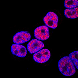
- Submitted by
- R&D Systems (provider)
- Main image
- Experimental details
- GATA-6 in HCT-116 Human Cell Line. GATA-6 was detected in immersion fixed HCT-116 human colorectal carcinoma cell line using Mouse Anti-Human GATA-6 Monoclonal Antibody (Catalog # MAB1700) at 10 µg/mL for 3 hours at room temperature. Cells were stained using the NorthernLights™ 557-conjugated Anti-Mouse IgG Secondary Antibody (red; Catalog # NL007) and counterstained with DAPI (blue). Specific staining was localized to nuclei. View our protocol for Fluorescent ICC Staining of Cells on Coverslips.

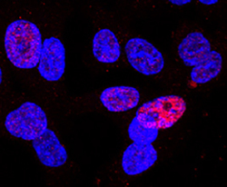
- Submitted by
- R&D Systems (provider)
- Main image

- Experimental details
- GATA-6 in HT-29 Human Cell Line. GATA-6 was detected in immersion fixed HT-29 human colon adenocarcinoma cell line using Mouse Anti-Human GATA-6 Monoclonal Antibody (Catalog # MAB1700) at 15 µg/mL for 3 hours at room temperature. Cells were stained using the NorthernLights™ 557-conjugated Anti-Mouse IgG Secondary Antibody (red; Catalog # NL007) and counterstained with DAPI (blue). Specific staining was localized to nuclei. View our protocol for Fluorescent ICC Staining of Cells on Coverslips.