Explore
 Validate
 Learn
LearnHPA039796
antibody from Atlas Antibodies
Targeting: CACNA1C
CACH2, CACN2, CACNL1A1, Cav1.2, CCHL1A1, LQT8, TS
 Immunohistochemistry
Antibody data
- Antibody Data
- Antigen structure
- References [0]
- Comments [0]
- Validations
- Immunohistochemistry [6]
Submit
Validation data
Reference
Comment
Report error
- Product number
- HPA039796 - Provider product page

- Provider
- Atlas Antibodies
- Proper citation
- Atlas Antibodies Cat#HPA039796, RRID:AB_10673019
- Product name
- Anti-CACNA1C
- Antibody type
- Polyclonal
- Description
- Affinity purified using the PrEST antigen as affinity ligand
- Reactivity
- Human
- Host
- Rabbit
- Conjugate
- Unconjugated
- Antigen sequence
QDETYEVKMNHDTEACSEPSLLSTEMLSYQDDENR
QLTLPEEDKRDIRQSPKRGFLRSASLGRRASFHLE
CLKRQKDRGGDISQKTVLPLHLVHHQALAVAGLS- Isotype
- IgG
- Vial size
- 100 µl
- Storage
- Store at +4°C for short term storage. Long time storage is recommended at -20°C.
No comments: Submit comment
Enhanced validation
Supportive validation
- Submitted by
- Atlas Antibodies (provider)
- Enhanced method
- Orthogonal validation
- Main image

- Experimental details
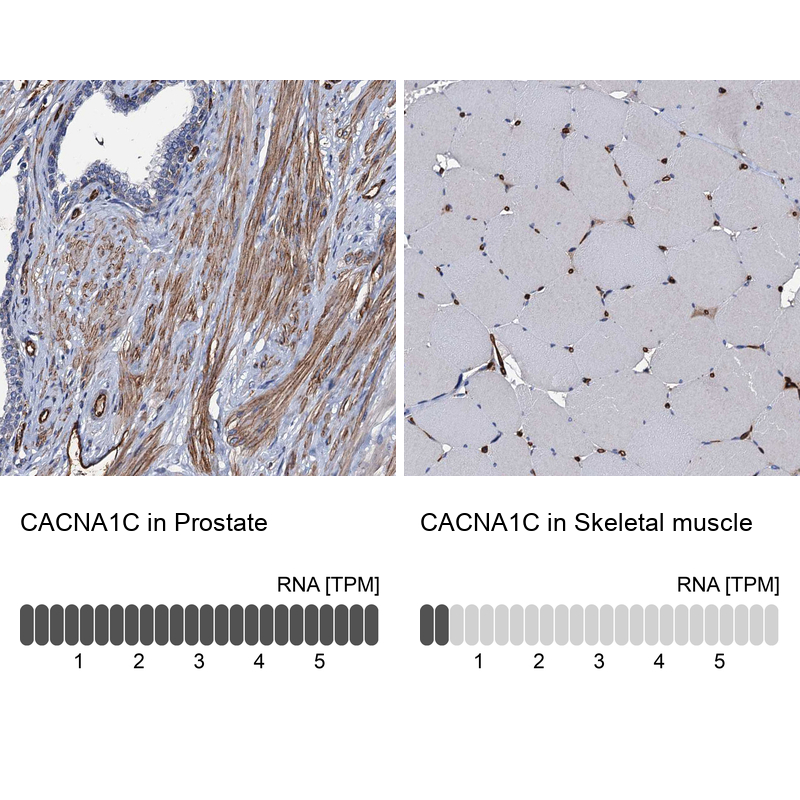
- Immunohistochemistry analysis in human prostate and skeletal muscle tissues using HPA039796 antibody. Corresponding CACNA1C RNA-seq data are presented for the same tissues.
- Sample type
- HUMAN
Supportive validation
- Submitted by
- Atlas Antibodies (provider)
- Main image

- Experimental details
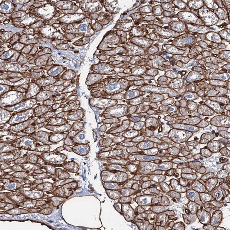
- Immunohistochemical staining of human heart muscle shows distinct membranous positivity in myocytes.
- Submitted by
- Atlas Antibodies (provider)
- Main image
- Experimental details
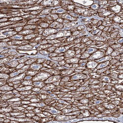
- Immunohistochemical staining of human heart shows strong positivity in plasma membrane of cardiomyocytes.
- Sample type
- HUMAN

- Submitted by
- Atlas Antibodies (provider)
- Main image
- Experimental details
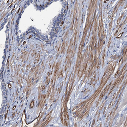
- Immunohistochemical staining of human prostate shows moderate positivity in plasma membrane in smooth muscle cells.
- Sample type
- HUMAN

- Submitted by
- Atlas Antibodies (provider)
- Main image
- Experimental details
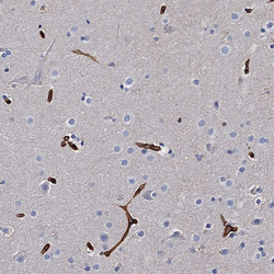
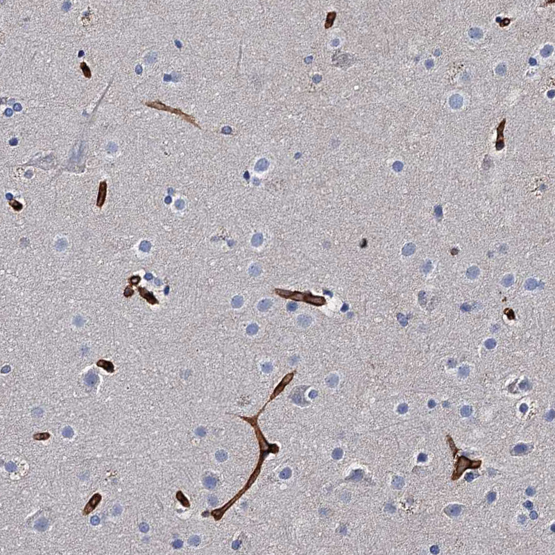
- Immunohistochemical staining of human cerebral cortex shows strong positivity in plasma membrane in endothelial cells.
- Sample type
- HUMAN
- Submitted by
- Atlas Antibodies (provider)
- Main image
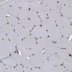
- Experimental details
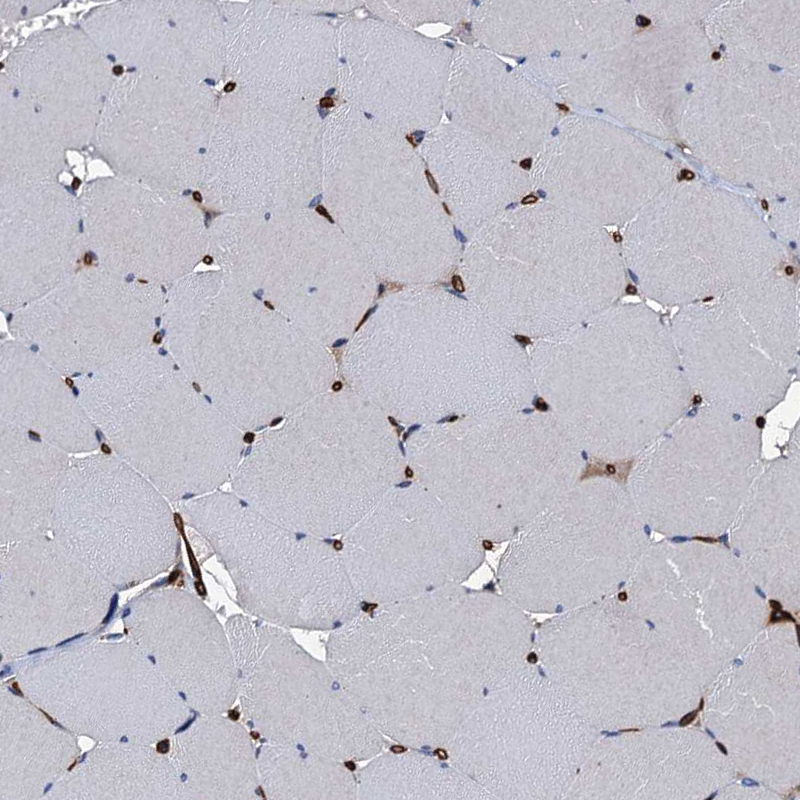
- Immunohistochemical staining of human skeletal muscle shows no positivity in striated muscle fibers as expected, while endothelium is strongly stained.
- Sample type
- HUMAN