Explore
 Validate
 Learn
Learn Western blot
Antibody data
- Antibody Data
- Antigen structure
- References [0]
- Comments [0]
- Validations
- Western blot [1]
- Immunohistochemistry [1]
- Flow cytometry [1]
Submit
Validation data
Reference
Comment
Report error
- Product number
- APC-150-200UL - Provider product page

- Provider
- Invitrogen Antibodies
- Product name
- KV1.5 (KCNA5) (extracellular) Polyclonal Antibody
- Antibody type
- Polyclonal
- Antigen
- Other
- Reactivity
- Human, Rat
- Host
- Rabbit
- Isotype
- IgG
- Vial size
- 200 µL
- Concentration
- 0.8 mg/mL
- Storage
- -20° C, Avoid Freeze/Thaw Cycles
No comments: Submit comment
Supportive validation
- Submitted by
- Invitrogen Antibodies (provider)
- Main image

- Experimental details
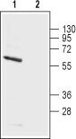
- Western blot analysis of rat brain membranes: - 1. Anti-KV1.5 (KCNA5) (extracellular) Antibody (#APC-150), (1:200). 2. Anti-KV1.5 (KCNA5) (extracellular) Antibody , preincubated with Kv1.5/KCNA5 (extracellular) Blocking Peptide (#BLP-PC150).
Supportive validation
- Submitted by
- Invitrogen Antibodies (provider)
- Main image
- Experimental details
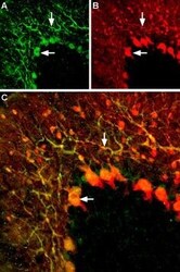
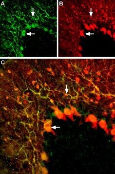
- Expression of KV1.5 channels in rat cerebellum - Immunohistochemical staining of rat cerebellum with Anti-KV1.5 (KCNA5) (extracellular) Antibody (#APC-150), (1:200). A. KV1.5 (green) appears in both the soma of Purkinje cells (horizontal arrows) and in Purkinje dendrites (vertical arrows). B. Neurons expressing gamma amino butyric acid (GABA) were labeled with mouse Anti-parvalbumin Antibody (red). C. Merge of the two images demonstrates partial colocalization (white arrows).
Supportive validation
- Submitted by
- Invitrogen Antibodies (provider)
- Main image
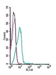
- Experimental details
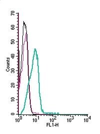
- Cell surface detection of KV1.5 by indirect flow cytometry in live intact human THP-1 monocytic leukemia cells: - (black line) cells. (red) Cells + goat- Anti-rabbit-FITC. (green) Cells + Anti-KV1.5 (KCNA5) (extracellular) Antibody (#APC-150), (5μg) + goat- Anti-rabbit-FITC.