 Explore
 Validate
 Learn
Learn Immunocytochemistry
Antibody data
- Antibody Data
- Antigen structure
- References [4]
- Comments [0]
- Validations
- Immunocytochemistry [1]
- Immunohistochemistry [1]
Submit
Validation data
Reference
Comment
Report error
- Product number
- HPA008773 - Provider product page

- Provider
- Atlas Antibodies
- Proper citation
- Atlas Antibodies Cat#HPA008773, RRID:AB_1078394
- Product name
- Anti-CA12
- Antibody type
- Polyclonal
- Reactivity
- Human
- Host
- Rabbit
- Conjugate
- Unconjugated
- Antigen sequence
TASNKSEGLAVLAVLIEMGSFNPSYDKIFSHLQHV
KYKGQEAFVPGFNIEELLPERTAEYYRYRGSLTTP
PCNPTVLWTVFRNPVQISQEQLLALETALYCTHMD
DPSPREMINNFRQVQ- Isotype
- IgG
- Vial size
- 100 µl
- Storage
- Store at +4°C for short term storage. Long time storage is recommended at -20°C.
Submitted references Differential expression of growth factor receptors and membrane-bound tumor markers for imaging in male and female breast cancer.
Immunophenotyping invasive breast cancer: paving the road for molecular imaging.
Noninvasive Detection of Breast Cancer Lymph Node Metastasis Using Carbonic Anhydrases IX and XII Targeted Imaging Probes
Gene expression signatures differentiate ovarian/peritoneal serous carcinoma from breast carcinoma in effusions
Vermeulen JF, Kornegoor R, van der Wall E, van der Groep P, van Diest PJ
PloS one 2013;8(1):e53353
PloS one 2013;8(1):e53353
Immunophenotyping invasive breast cancer: paving the road for molecular imaging.
Vermeulen JF, van Brussel AS, van der Groep P, Morsink FH, Bult P, van der Wall E, van Diest PJ
BMC cancer 2012 Jun 13;12:240
BMC cancer 2012 Jun 13;12:240
Noninvasive Detection of Breast Cancer Lymph Node Metastasis Using Carbonic Anhydrases IX and XII Targeted Imaging Probes
Tafreshi N, Bui M, Bishop K, Lloyd M, Enkemann S, Lopez A, Abrahams D, Carter B, Vagner J, Grobmyer S, Gillies R, Morse D
Clinical Cancer Research 2012 January;18(1):207-219
Clinical Cancer Research 2012 January;18(1):207-219
Gene expression signatures differentiate ovarian/peritoneal serous carcinoma from breast carcinoma in effusions
Davidson B, Stavnes H, Holth A, Chen X, Yang Y, Shih I, Wang T
Journal of Cellular and Molecular Medicine 2010 January
Journal of Cellular and Molecular Medicine 2010 January
No comments: Submit comment
Supportive validation
- Submitted by
- Atlas Antibodies (provider)
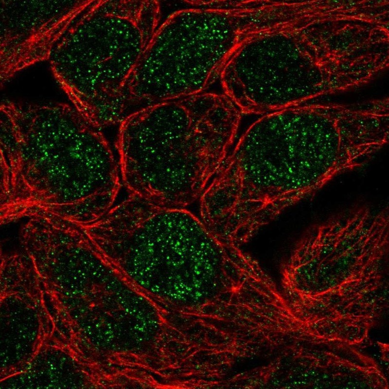
- Main image

- Experimental details
- Immunofluorescent staining of human cell line RT4 shows localization to nucleus.
- Sample type
- HUMAN
Supportive validation
- Submitted by
- Atlas Antibodies (provider)
- Main image
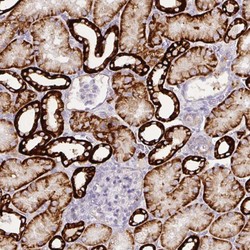
- Experimental details
- Immunohistochemical staining of human kidney shows strong cytoplasmic and membranous positivity in cells in tubules.
- Sample type
- HUMAN
