Explore
 Validate
 Learn
Learn Western blot
 Immunoprecipitation
Antibody data
- Antibody Data
- Antigen structure
- References [3]
- Comments [0]
- Validations
- Western blot [6]
- Immunoprecipitation [1]
Submit
Validation data
Reference
Comment
Report error
- Product number
- GTX129528 - Provider product page

- Provider
- GeneTex
- Product name
- VPS34 antibody
- Antibody type
- Polyclonal
- Reactivity
- Human
- Host
- Rabbit
Submitted references Dual Inhibition of PIK3C3 and FGFR as a New Therapeutic Approach to Treat Bladder Cancer.
GSK3B-mediated phosphorylation of MCL1 regulates axonal autophagy to promote Wallerian degeneration.
VPS34 stimulation of p62 phosphorylation for cancer progression.
Chen CH, Changou CA, Hsieh TH, Lee YC, Chu CY, Hsu KC, Wang HC, Lin YC, Lo YN, Liu YR, Liou JP, Yen Y
Clinical cancer research : an official journal of the American Association for Cancer Research 2018 Mar 1;24(5):1176-1189
Clinical cancer research : an official journal of the American Association for Cancer Research 2018 Mar 1;24(5):1176-1189
GSK3B-mediated phosphorylation of MCL1 regulates axonal autophagy to promote Wallerian degeneration.
Wakatsuki S, Tokunaga S, Shibata M, Araki T
The Journal of cell biology 2017 Feb;216(2):477-493
The Journal of cell biology 2017 Feb;216(2):477-493
VPS34 stimulation of p62 phosphorylation for cancer progression.
Jiang X, Bao Y, Liu H, Kou X, Zhang Z, Sun F, Qian Z, Lin Z, Li X, Liu X, Jiang L, Yang Y
Oncogene 2017 Dec 14;36(50):6850-6862
Oncogene 2017 Dec 14;36(50):6850-6862
No comments: Submit comment
Enhanced validation
Supportive validation
- Submitted by
- GeneTex (provider)
- Enhanced method
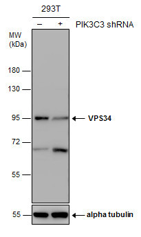
- Genetic validation
- Main image

- Experimental details
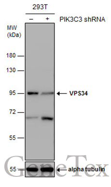
- Non-transfected (¡V) and transfected (+) 293T whole cell extracts (30 ?g) were separated by 7.5% SDS-PAGE, and the membrane was blotted with VPS34 antibody (GTX129528) diluted at 1:1000.
Supportive validation
- Submitted by
- GeneTex (provider)
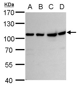
- Main image

- Experimental details
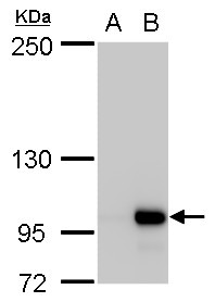
- VPS34 antibody detects VPS34 protein by western blot analysis.A. 30 £gg 293T whole cell lysate/extract B. 30 £gg A431 whole cell lysate/extract C. 30 £gg HeLa whole cell lysate/extract D. 30 £gg HepG2 whole cell lysate/extract7.5 % SDS-PAGEVPS34 antibody (GTX129528) dilution: 1:1000
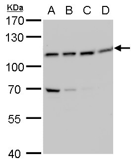
- Submitted by
- GeneTex (provider)
- Main image

- Experimental details
- VPS34 antibody detects VPS34 protein by western blot analysis.A. 30 £gg U87-MG whole cell lysate/extract B. 30 £gg SK-N-SH whole cell lysate/extract C. 30 £gg IMR32 whole cell lysate/extract D. 30 £gg SK-N-AS whole cell lysate/extract7.5 % SDS-PAGEVPS34 antibody (GTX129528) dilution: 1:1000
- Submitted by
- GeneTex (provider)
- Main image

- Experimental details
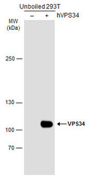
- VPS34 antibody detects VPS34 protein by western blot analysis.A. 30 £gg 293T whole cell lysate/extract B. 30 £gg whole cell lysate/extract of human VPS34-transfected 293T cells5 % SDS-PAGEVPS34 antibody (GTX129528) dilution: 1:5000
- Submitted by
- GeneTex (provider)
- Main image

- Experimental details
- Non-transfected (¡V) and transfected (+) 293T whole cell extracts (30 ?g) were separated by 7.5% SDS-PAGE, and the membrane was blotted with VPS34 antibody (GTX129528) diluted at 1:1000.
- Submitted by
- GeneTex (provider)
- Main image
- Experimental details
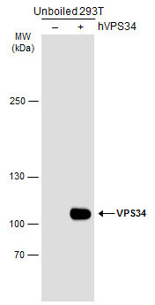
- Non-transfected (¡V) and transfected (+) unboiled 293T whole cell extracts (30 ?g) were separated by 5% SDS-PAGE, and the membrane was blotted with VPS34 antibody (GTX129528) diluted at 1:5000. The HRP-conjugated anti-rabbit IgG antibody (GTX213110-01) was used to detect the primary antibody.
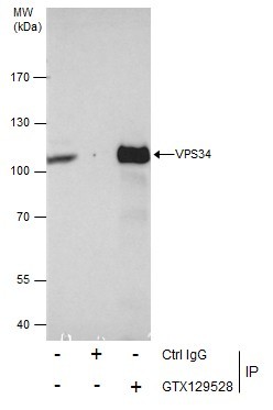
Supportive validation
- Submitted by
- GeneTex (provider)
- Main image

- Experimental details
- Immunoprecipitation of VPS34 protein from HeLa whole cell extracts using 5 £gg of VPS34 antibody (GTX129528).Western blot analysis was performed using VPS34 antibody (GTX129528).EasyBlot anti-Rabbit IgG (GTX221666-01) was used as a secondary reagent.