Explore
 Validate
 Learn
LearnPA5-26778
antibody from Invitrogen Antibodies
Targeting: ATP5F1A
ATP5A, ATP5A1, ATP5AL2, ATPM, hATP1, OMR, ORM
 Western blot
Antibody data
- Antibody Data
- Antigen structure
- References [1]
- Comments [0]
- Validations
- Western blot [3]
- Immunocytochemistry [1]
- Immunohistochemistry [1]
- Other assay [1]
Submit
Validation data
Reference
Comment
Report error
- Product number
- PA5-26778 - Provider product page

- Provider
- Invitrogen Antibodies
- Product name
- ATP5A1 Polyclonal Antibody
- Antibody type
- Polyclonal
- Antigen
- Synthetic peptide
- Description
- This antibody is predicted to react with bovine, mouse, porcine and rat based on sequence homology.
- Reactivity
- Human
- Host
- Rabbit
- Isotype
- IgG
- Vial size
- 400 µL
- Concentration
- 0.48 mg/mL
- Storage
- Store at 4°C short term. For long term storage, store at -20°C, avoiding freeze/thaw cycles.
Submitted references pH sensing in skin tumors: Methods to study the involvement of GPCRs, acid-sensing ion channels and transient receptor potential vanilloid channels.
Stolwijk JA, Sauer L, Ackermann K, Nassios A, Aung T, Haerteis S, Bäumner AJ, Wegener J, Schreml S
Experimental dermatology 2020 Nov;29(11):1055-1061
Experimental dermatology 2020 Nov;29(11):1055-1061
No comments: Submit comment
Supportive validation
- Submitted by
- Invitrogen Antibodies (provider)
- Main image

- Experimental details
- Western blot analysis using an ATP5A1 polyclonal antibody (Product # PA5-26778) in WiDr, NCI-H460, MDA-MB231 cell lysates (35 µg per lane).

- Submitted by
- Invitrogen Antibodies (provider)
- Main image

- Experimental details
- Western blot analysis using an ATP5A1 polyclonal antibody (Product # PA5-26778) in WiDr, NCI-H460, MDA-MB231 cell lysates (35 µg per lane).

- Submitted by
- Invitrogen Antibodies (provider)
- Main image

- Experimental details
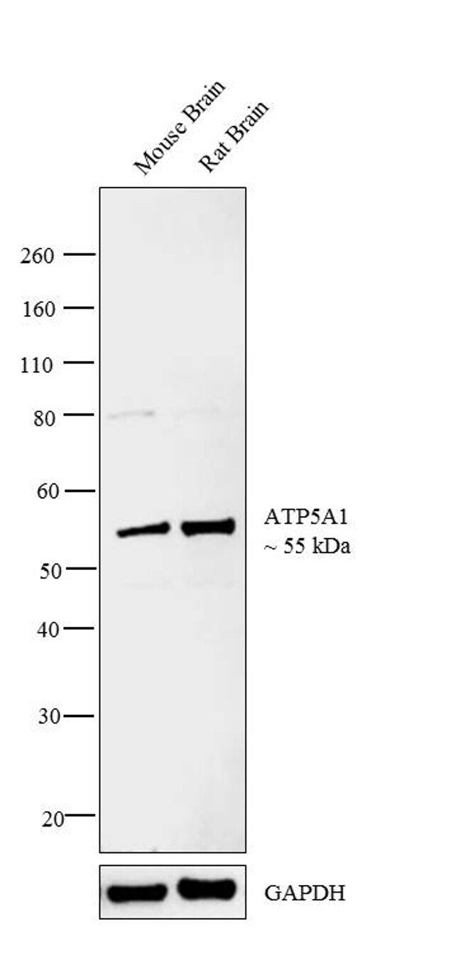
- Western blot analysis was performed on tissue extracts (30 µg lysate) of Mouse Brain (Lane 1) and Rat Brain (Lane 2). The blot was probed with Anti-ATP5A1 Polyclonal Antibody (Product # PA5-26778, 1:500 dilution) and detected by chemiluminescence using Goat anti-Rabbit IgG (H+L) Superclonal™ Secondary Antibody, HRP conjugate (Product # A27036, 0.25 µg/mL, 1:4000 dilution). A 55 kDa band corresponding to ATP5A1 was detected across the tissues tested.
Supportive validation
- Submitted by
- Invitrogen Antibodies (provider)
- Main image

- Experimental details
- Immunofluorescence analysis of ATP5A1 was performed using 70% confluent log phase HeLa cells. The cells were fixed with 4% paraformaldehyde for 10 minutes, permeabilized with 0.1% Triton™ X-100 for 15 minutes, and blocked with 1% BSA for 1 hour at room temperature. The cells were labeled with ATP5A1 Polyclonal Antibody (Product # PA5-26778) at 1:250 dilution in 0.1% BSA, incubated at 4 degree Celsius overnight and then labeled with Goat anti-Rabbit IgG (H+L) Superclonal™ Secondary Antibody, Alexa Fluor® 488 conjugate (Product # A27034) at a dilution of 1:2000 for 45 minutes at room temperature (Panel a: green). Nuclei (Panel b: blue) were stained with ProLong™ Diamond Antifade Mountant with DAPI (Product # P36962). F-actin (Panel c: red) was stained with Rhodamine Phalloidin (Product # R415, 1:300). Panel d represents the merged image showing mitochondrial localization. Panel e represents control cells with no primary antibody to assess background. The images were captured at 60X magnification.

Supportive validation
- Submitted by
- Invitrogen Antibodies (provider)
- Main image

- Experimental details
- Immunohistochemistry analysis in formalin-fixed, paraffin-embedded lung carcinoma using an ATP5A1 polyclonal antibody (Product # PA5-26778), followed by HRP-conjugated secondary antibody and DAB staining.

Supportive validation
- Submitted by
- Invitrogen Antibodies (provider)
- Main image
- Experimental details
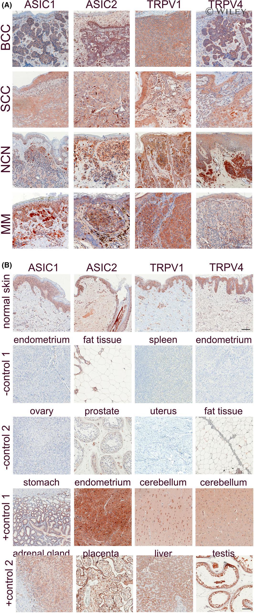
- 1 FIGURE Immunohistochemistry of ASIC1/2 and TRPV1/4: (A) Representative samples of the most common skin tumors, that is basal cell carcinoma (BCC), squamous cell carcinoma (SCC), naevus cell naevus (NCN) and melanoma (MM) were stained for ASIC1, ASIC2, TRPV1 or TRPV4, according to the protocol as described in Nassios et al [ 36 ] and using the following primary antibodies: ASIC1: PA5-26778, ASIC2: PA5-26222, TRPV1: LS-B12677 (Thermo Fisher Scientific Inc, Waltham, MA, USA), TRPV4: ab21912 (abcam, Cambridge, UK). B, Normal skin (first row) and control tissues (second to fifth row) were stained for ASIC1, ASIC2, TRPV1 or TRPV4. Negative (second and third row) and positive (fourth and fifth row) controls were selected according to expression analyses published at the human protein atlas ( https://www.proteinatlas.org/ ). Scale bars: 100 um. Compared to epidermis, BCC showed strong expression of the four pH-sensitive proteins. In contrast, SCC showed a quite similar expression of all pH-sensitive proteins in comparison to normal epidermis, markedly weaker than BCC. For NCN and MM, epidermal and dermal expression was quite different. NCN exhibit strong expression of ASIC1/2 in the more superficial portion, while the intensity decreases in dermal melanocytes. However, TRPV1/4 seems to be uniformly expressed even in deeper tissue layers. In MM, ASIC1/2 and also TRPV1 are also expressed throughout the whole tumor. In contrast, TRPV4 staining showed only weak expression in MM, which