Antibody data
- Antibody Data
- Antigen structure
- References [3]
- Comments [0]
- Validations
- Western blot [1]
Submit
Validation data
Reference
Comment
Report error
- Product number
- PAB10080 - Provider product page

- Provider
- Abnova Corporation
- Proper citation
- Abnova Corporation Cat#PAB10080, RRID:AB_1679622
- Product name
- ING4 polyclonal antibody
- Antibody type
- Polyclonal
- Description
- Goat polyclonal antibody raised against synthetic peptide of ING4.
- Storage
- Store at 4°C. For long term storage store at -20°C.Aliquot to avoid repeated freezing and thawing.
Submitted references A screen for genes that suppress loss of contact inhibition: identification of ING4 as a candidate tumor suppressor gene in human cancer.
The candidate tumour suppressor protein ING4 regulates brain tumour growth and angiogenesis.
ING4 induces G2/M cell cycle arrest and enhances the chemosensitivity to DNA-damage agents in HepG2 cells.
Kim S, Chin K, Gray JW, Bishop JM
Proceedings of the National Academy of Sciences of the United States of America 2004 Nov 16;101(46):16251-6
Proceedings of the National Academy of Sciences of the United States of America 2004 Nov 16;101(46):16251-6
The candidate tumour suppressor protein ING4 regulates brain tumour growth and angiogenesis.
Garkavtsev I, Kozin SV, Chernova O, Xu L, Winkler F, Brown E, Barnett GH, Jain RK
Nature 2004 Mar 18;428(6980):328-32
Nature 2004 Mar 18;428(6980):328-32
ING4 induces G2/M cell cycle arrest and enhances the chemosensitivity to DNA-damage agents in HepG2 cells.
Zhang X, Xu LS, Wang ZQ, Wang KS, Li N, Cheng ZH, Huang SZ, Wei DZ, Han ZG
FEBS letters 2004 Jul 16;570(1-3):7-12
FEBS letters 2004 Jul 16;570(1-3):7-12
No comments: Submit comment
Supportive validation
- Submitted by
- Abnova Corporation (provider)
- Main image

- Experimental details
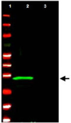
- ING4 polyclonal antibody (Cat # PAB10080) detects ING4 protein by western blot.This antibody was used at 1.0 ug/mL to detect ING4 (Lane 2) present in a U-2 OS whole cell lysate over expressing the protein.A control lysate (Lane 3) shows no background staining.Comparison to MW markers (Lane 1) indicates detection of a single band at ~29 KDa corresponding to ING4.A 4-20% TRIS-glycine gradient gel was used to separate the protein by SDS-PAGE under reducing conditions.The protein was transferred to nitrocellulose using standard methods.After blocking using 5% non-fat dry milk in PBS, the membrane was probed with the primary antibody overnight at 4°C followed by washes and reaction with a 1:20,000 dilution of IRDye™800 conjugated Rb-a-Goat IgG [H&L] for 45 min at room temperature.LICOR's Odyssey® Infrared Imaging System was used to scan and process the image.
 Explore
 Validate
 Learn
Learn Western blot