 Explore
 Validate
 Learn
Learn Western blot
Antibody data
- Antibody Data
- Antigen structure
- References [3]
- Comments [0]
- Validations
- Western blot [2]
- Immunocytochemistry [1]
- Immunohistochemistry [1]
Submit
Validation data
Reference
Comment
Report error
- Product number
- AF2517 - Provider product page

- Provider
- R&D Systems
- Product name
- Mouse/Rat PDX-1/IPF1 Antibody
- Antibody type
- Polyclonal
- Description
- Antigen Affinity-purified. Detects mouse PDX-1/IPF1 in direct ELISAs and Western blots.
- Reactivity
- Mouse, Rat
- Host
- Goat
- Conjugate
- Unconjugated
- Antigen sequence
P52946- Isotype
- IgG
- Vial size
- 100 ug
- Concentration
- LYOPH
- Storage
- Use a manual defrost freezer and avoid repeated freeze-thaw cycles. 12 months from date of receipt, -20 to -70 °C as supplied. 1 month, 2 to 8 °C under sterile conditions after reconstitution. 6 months, -20 to -70 °C under sterile conditions after reconstitution.
Submitted references Characterization of induced tissue-specific stem cells from pancreas by a synthetic self-replicative RNA.
Per-Arnt-Sim kinase regulates pancreatic duodenal homeobox-1 protein stability via phosphorylation of glycogen synthase kinase 3β in pancreatic β-cells.
Self-renewal of embryonic-stem-cell-derived progenitors by organ-matched mesenchyme.
Miyagi-Shiohira C, Nakashima Y, Kobayashi N, Saitoh I, Watanabe M, Noguchi H
Scientific reports 2018 Aug 17;8(1):12341
Scientific reports 2018 Aug 17;8(1):12341
Per-Arnt-Sim kinase regulates pancreatic duodenal homeobox-1 protein stability via phosphorylation of glycogen synthase kinase 3β in pancreatic β-cells.
Semache M, Zarrouki B, Fontés G, Fogarty S, Kikani C, Chawki MB, Rutter J, Poitout V
The Journal of biological chemistry 2013 Aug 23;288(34):24825-33
The Journal of biological chemistry 2013 Aug 23;288(34):24825-33
Self-renewal of embryonic-stem-cell-derived progenitors by organ-matched mesenchyme.
Sneddon JB, Borowiak M, Melton DA
Nature 2012 Nov 29;491(7426):765-8
Nature 2012 Nov 29;491(7426):765-8
No comments: Submit comment
Supportive validation
- Submitted by
- R&D Systems (provider)
- Main image
- Experimental details
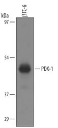
- Detection of Mouse/Rat PDX-1/IPF1 by Western Blot. Western blot shows lysates of beta TC-6 mouse beta cell insulinoma cell line. PVDF membrane was probed with 1 µg/mL of Goat Anti-Mouse/Rat PDX-1/IPF1 Antigen Affinity-purified Polyclonal Antibody (Catalog # AF2517) followed by HRP-conjugated Anti-Goat IgG Secondary Antibody (Catalog # HAF019). A specific band was detected for PDX-1/IPF1 at approximately 45 kDa (as indicated). This experiment was conducted under reducing conditions and using Immunoblot Buffer Group 8.

- Submitted by
- R&D Systems (provider)
- Main image

- Experimental details
- Detection of Mouse PDX-1/IPF1 by Simple WesternTM. Simple Western lane view shows lysates of beta TC-6 mouse beta cell insulinoma cell line, loaded at 0.2 mg/mL. A specific band was detected for PDX-1/IPF1 at approximately 48 kDa (as indicated) using 10 µg/mL of Goat Anti-Mouse/Rat PDX-1/IPF1 Antigen Affinity-purified Polyclonal Antibody (Catalog # AF2517) followed by 1:50 dilution of HRP-conjugated Anti-Goat IgG Secondary Antibody (Catalog # HAF109). This experiment was conducted under reducing conditions and using the 12-230 kDa separation system.

Supportive validation
- Submitted by
- R&D Systems (provider)
- Main image

- Experimental details
- PDX-1/IPF1 in beta TC-6 Mouse Cell Line. PDX-1/IPF1 was detected in immersion fixed beta TC-6 mouse beta cell insulinoma cell line using Goat Anti-Mouse/Rat PDX-1/IPF1 Antigen Affinity-purified Polyclonal Antibody (Catalog # AF2517) at 10 µg/mL for 3 hours at room temperature. Cells were stained using the NorthernLights™ 557-conjugated Anti-Goat IgG Secondary Antibody (red; Catalog # NL001) and counterstained with DAPI (blue). Specific staining was localized to the nucleus. View our protocol for Fluorescent ICC Staining of Cells on Coverslips.

Supportive validation
- Submitted by
- R&D Systems (provider)
- Main image
- Experimental details
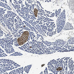
- PDX-1/IPF1 in Mouse Pancreas. PDX-1/IPF1 was detected in perfusion fixed frozen sections of mouse pancreas using Goat Anti-Mouse/Rat PDX-1/IPF1 Antigen Affinity-purified Polyclonal Antibody (Catalog # AF2517) at 3 µg/mL for 1 hour at room temperature followed by incubation with the Anti-Goat IgG VisUCyte™ HRP Polymer Antibody (Catalog # VC004). Tissue was stained using DAB (brown) and counterstained with hematoxylin (blue). View our protocol for IHC Staining with VisUCyte HRP Polymer Detection Reagents.
