 Explore
 Validate
 Learn
LearnPA5-32046
antibody from Invitrogen Antibodies
Targeting: STUB1
CHIP, HSPABP2, NY-CO-7, SDCCAG7, UBOX1
 Western blot
Antibody data
- Antibody Data
- Antigen structure
- References [2]
- Comments [0]
- Validations
- Western blot [7]
- Immunohistochemistry [1]
- Other assay [1]
Submit
Validation data
Reference
Comment
Report error
- Product number
- PA5-32046 - Provider product page

- Provider
- Invitrogen Antibodies
- Product name
- STUB1 Polyclonal Antibody
- Antibody type
- Polyclonal
- Antigen
- Recombinant protein fragment
- Description
- Recommended positive controls: 293T, A431, Raji, BT-474, HCC1937, Hs578T, MCF7, MDA-MB-231, MDA-MB-361, MDA-MB-453, T-47D, ZR-75-1, HeLa, U2OS.
- Concentration
- 1 mg/mL
Submitted references Disruption of the menin-MLL interaction triggers menin protein degradation via ubiquitin-proteasome pathway.
Hsp70 and Hsp40 inhibit an inter-domain interaction necessary for transcriptional activity in the androgen receptor.
Wu Y, Doepner M, Hojnacki T, Feng Z, Katona BW, He X, Ma J, Cao Y, Busino L, Zhou F, Hua X
American journal of cancer research 2019;9(8):1682-1694
American journal of cancer research 2019;9(8):1682-1694
Hsp70 and Hsp40 inhibit an inter-domain interaction necessary for transcriptional activity in the androgen receptor.
Eftekharzadeh B, Banduseela VC, Chiesa G, Martínez-Cristóbal P, Rauch JN, Nath SR, Schwarz DMC, Shao H, Marin-Argany M, Di Sanza C, Giorgetti E, Yu Z, Pierattelli R, Felli IC, Brun-Heath I, García J, Nebreda ÁR, Gestwicki JE, Lieberman AP, Salvatella X
Nature communications 2019 Aug 8;10(1):3562
Nature communications 2019 Aug 8;10(1):3562
No comments: Submit comment
Supportive validation
- Submitted by
- Invitrogen Antibodies (provider)
- Main image

- Experimental details
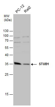
- Western blot analysis of STUB1 in Various whole cell extracts (30 µg). Samples were separated by 12% SDS-PAGE and the membrane was probed with STUB1 Polyclonal antibody (Product # PA5-32046) at a dilution of 1:10000.

- Submitted by
- Invitrogen Antibodies (provider)
- Main image

- Experimental details
- Western Blot using STUB1 Polyclonal Antibody (Product # PA5-32046). Various whole cell extracts (30 µg) were separated by 12% SDS-PAGE, and the membrane was blotted with STUB1 Polyclonal Antibody (Product # PA5-32046) diluted at 1:10,000.

- Submitted by
- Invitrogen Antibodies (provider)
- Main image

- Experimental details
- Western Blot using STUB1 Polyclonal Antibody (Product # PA5-32046). Sample (30 µg of whole cell lysate). Lane A: 293T. 10% SDS PAGE. STUB1 Polyclonal Antibody (Product # PA5-32046) diluted at 1:10,000.

- Submitted by
- Invitrogen Antibodies (provider)
- Main image

- Experimental details
- Western Blot using STUB1 Polyclonal Antibody (Product # PA5-32046). Various whole cell extracts (30 µg) were separated by 12% SDS-PAGE, and the membrane was blotted with STUB1 Polyclonal Antibody (Product # PA5-32046) diluted at 1:10,000.

- Submitted by
- Invitrogen Antibodies (provider)
- Main image

- Experimental details
- Western Blot using STUB1 Polyclonal Antibody (Product # PA5-32046). Various whole cell extracts (30 µg) were separated by 12% SDS-PAGE, and the membrane was blotted with STUB1 Polyclonal Antibody (Product # PA5-32046) diluted at 1:10,000.
- Submitted by
- Invitrogen Antibodies (provider)
- Main image

- Experimental details
- Western blot was performed using Anti-E3 ubiquitin-protein ligase CHIP (STUB1) Polyclonal Antibody (Product # PA5-32046) on whole cell extracts (30 µg lysate) of HEK-293 (Lane 1), MCF7 (Lane 2), HeLa (Lane 3), LNCaP (Lane 4), Jurkat (Lane 5) and Hep G2 (Lane 6) and 35 kDa band corresponding to STUB1 was observed except in Jurkat and Hep G2 along with an uncharacterized band (*) at ~ 110 kDa. Resolved proteins were then transferred onto a nitrocellulose membrane (Product # IB23001) by iBlot® 2 Dry Blotting System (Product # IB21001). The blot was probed with the primary antibody (1:10,000 dilution) and detected by Goat anti-Rabbit IgG (H+L) Superclonal™ Recombinant Secondary Antibody, HRP conjugate (Product # A27036, 0.25 µg/ml, 1:4000 dilution) using the iBright FL 1000 (Product # A32752). Chemiluminescent detection was performed using Novex® ECL Chemiluminescent Substrate Reagent Kit (Product # WP20005).

Supportive validation
- Submitted by
- Invitrogen Antibodies (provider)
- Main image

- Experimental details
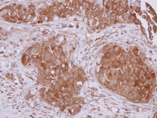
- Immunohistochemical analysis of paraffin-embedded human breast cancer, using STUB1 (Product # PA5-32046) antibody at 1:500 dilution. Antigen Retrieval: Citrate buffer, pH 6.0, 15 min.
Supportive validation
- Submitted by
- Invitrogen Antibodies (provider)
- Main image

- Experimental details
- Fig. 5 Hsp70-targeted small molecules increase ubiquitination and degradation of polyQ-AR. a PC12 cells were transiently transfected to express HA-ubiquitin and CHIP, and AR112Q expression was induced for 48 h. Cells were treated with the Hsp70 modulator, JG-98 (0.5 muM) for the last 24 h and with 10 muM MG132 for the last 16 h. AR112Q was immuno-precipitated from lysates and then probed for ubiquitin (HA). Left: input. Middle: pull down. Right: quantification from three independent experiments. Data are mean +- S.E.M. from three independent experiments. * p < 0.05 by two-tailed t -test. b JG-98 promotes AR112Q degradation by the proteasome. PC12 cells were induced to express AR112Q for 48 h in the presence of R1881 (10 nM) and JG-98 (0.5 or 1.0 muM). Indicated samples were treated with lactacystine (10 muM) for the final 16 h. AR was detected by western blot. GAPDH controls for loading. c JG-98 does not induce a stress response. PC12 cells were induced to express AR112Q in the presence of R1881 (10 nM) for 48 h in the presence of 17-AAG (5 muM), JG-98 (0.5 muM) or vehicle control. Cell lysates were probed for Hsp25, Hsp40, Hsp70, Hsp90, Akt, and ERK1/2. GAPDH controls for loading. d PC12 cells were induced to express AR112Q in the presence of R1881 for 48 h, and treated with 17-AAG (1 uM) and/or JG-98 (1 uM), as indicated. Lysates were analyzed for AR by filter trap assay. Data are mean +- S.E.M. from three independent experiments. * p < 0.05, ** p < 0.01, *** p < 0.001 by o
