Antibody data
- Antibody Data
- Antigen structure
- References [1]
- Comments [0]
- Validations
- Western blot [1]
- Immunocytochemistry [1]
- Immunohistochemistry [2]
- Flow cytometry [1]
Submit
Validation data
Reference
Comment
Report error
- Product number
- GTX20262 - Provider product page

- Provider
- GeneTex
- Product name
- EAAT1 antibody
- Antibody type
- Polyclonal
- Reactivity
- Human, Mouse, Rat, Bovine
- Host
- Rabbit
Submitted references Aspartate is a limiting metabolite for cancer cell proliferation under hypoxia and in tumours.
Garcia-Bermudez J, Baudrier L, La K, Zhu XG, Fidelin J, Sviderskiy VO, Papagiannakopoulos T, Molina H, Snuderl M, Lewis CA, Possemato RL, Birsoy K
Nature cell biology 2018 Jul;20(7):775-781
Nature cell biology 2018 Jul;20(7):775-781
No comments: Submit comment
Supportive validation
- Submitted by
- GeneTex (provider)
- Main image

- Experimental details
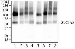
- Western Blot: SLC1A3 Antibody - Western blot analysis of human brain tissue (1), human brain membrane tissue (2), human hippocampus tissue (3), bovine brain (4), rat brain tissue (5), rat brain membrane tissue (6), mouse brain tissue (7), and mouse membrane tissue (8) using SLC1A3 antibody at 2 μg/ml.
Supportive validation
- Submitted by
- GeneTex (provider)
- Main image

- Experimental details
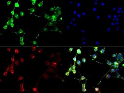
- Immunocytochemistry/Immunofluorescence: SLC1A3 Antibody - SLC1A3 antibody was tested at (1:250) in Neuro2A cells with DyLight 488 (green). Nuclei and alpha-tubulin were counterstained with DAPI (blue) and Dylight 550 (red). Image objective 40x.
Supportive validation
- Submitted by
- GeneTex (provider)
- Main image

- Experimental details
- Immunohistochemistry-Paraffin: SLC1A3 Antibody - IHC-P analysis of SLC1A3 on mouse brain.
- Submitted by
- GeneTex (provider)
- Main image

- Experimental details
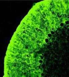
- Immunohistochemistry: SLC1A3 Antibody - IHC staining of SLC1A3 in rat cerebellum sections
Supportive validation
- Submitted by
- GeneTex (provider)
- Main image

- Experimental details
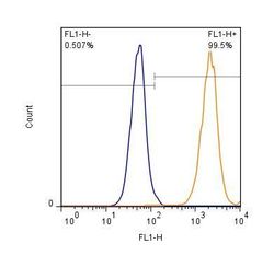
- Flow (Intracellular): EAAT1/GLAST-1/SLC1A3 Antibody - Intracellular staining of HEK293 cells (1 x 10^6 cells/ml) with SLC1A3 antibody (orange) stained at a dilution of 1:500. Detected with a Dylight 488 secondary antibody. Shown with the secondary control (blue).
 Explore
 Validate
 Learn
Learn Western blot
 ELISA
ELISA