Antibody data
- Antibody Data
- Antigen structure
- References [1]
- Comments [0]
- Validations
- Western blot [2]
- Immunocytochemistry [1]
- Other assay [1]
Submit
Validation data
Reference
Comment
Report error
- Product number
- PA5-106771 - Provider product page

- Provider
- Invitrogen Antibodies
- Product name
- CaV3.2 Polyclonal Antibody
- Antibody type
- Polyclonal
- Antigen
- Synthetic peptide
- Description
- Antibody detects endogenous levels of total CACNA1H.
- Reactivity
- Human, Mouse, Rat
- Host
- Rabbit
- Isotype
- IgG
- Vial size
- 100 µL
- Concentration
- 1 mg/mL
- Storage
- -20°C
Submitted references Evidence for a Physiological Role of T-Type Ca Channels in Ventricular Cardiomyocytes of Adult Mice.
Marksteiner J, Ebner J, Salzer I, Lilliu E, Hackl B, Todt H, Kubista H, Hallström S, Koenig X, Hilber K
Membranes 2022 May 28;12(6)
Membranes 2022 May 28;12(6)
No comments: Submit comment
Supportive validation
- Submitted by
- Invitrogen Antibodies (provider)
- Main image

- Experimental details
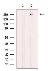
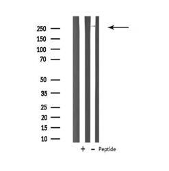
- Western blot analysis of CaV3.2 in A549 cell lysate (left lane: treated with blocking peptide). Samples were incubated with CaV3.2 polyclonal antibody (Product # PA5-106771).
- Submitted by
- Invitrogen Antibodies (provider)
- Main image

- Experimental details
- Western blot analysis of CaV3.2 in A549 cells. Samples were incubated with CaV3.2 polyclonal antibody (Product # PA5-106771).
Supportive validation
- Submitted by
- Invitrogen Antibodies (provider)
- Main image

- Experimental details
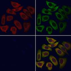
- Immunofluorescent analysis of CaV3.2 in HeLa cells. Samples were fixed with paraformaldehyde, permeabilized with 0.1% Triton X-100, blocked with 10% serum (45 min at 25°C), incubated with mouse anti-beta tubulin and CaV3.2 polyclonal antibody (Product # PA5-106771) using a dilution of 1:200 (1 hr, 37°C), and followed by goat anti-rabbit IgG Alexa Fluor 594 (red) and goat anti-mouse IgG Alexa Fluor 488 (green).
Supportive validation
- Submitted by
- Invitrogen Antibodies (provider)
- Main image

- Experimental details
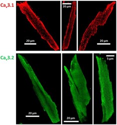
- Immunostaining of T-type Ca channels in ventricular cardiomyocytes isolated from adult wt mice. Cav3.1 ( top ) channel expression and localization was detected using a selective anti-Cav3.1 antibody (anti-CACNA1G, #ACC-021, source: rabbit, Alomone Labs, 1:500). Cav3.2 ( bottom ) was detected with the selective anti-Cav3.2 antibodies (anti-CACNA1H, #ACC-025, source: rabbit, Alomone Labs, 1:1000; left and right cell; or anti-Cav3.2 polyclonal, #PA5-106771, source: rabbit, Invitrogen, 1:200; middle cell). Secondary antibody: Alexa Fluor 488, #A11008, goat anti-rabbit, Invitrogen, 1:500.
 Explore
 Validate
 Learn
Learn Western blot