 Explore
 Validate
 Learn
Learn Western blot
Antibody data
- Antibody Data
- Antigen structure
- References [0]
- Comments [0]
- Validations
- Western blot [1]
- Immunocytochemistry [2]
- Immunohistochemistry [2]
Submit
Validation data
Reference
Comment
Report error
- Product number
- 102-PA36 - Provider product page

- Provider
- ReliaTech GmbH
- Product name
- ccbe1
- Antibody type
- Polyclonal
- Antigen
- recombinant human ccbe1
- Description
- antibody Protein-A purified from serum
- Reactivity
- Human
- Host
- Rabbit
- Antigen sequence
YREEPEDGDREICSESKIATTKYPCLKSSGELTTC
YRKKCCKGYKFVLGQCIPEDYDVCAEAPCEQQCTD
NFGRVLCTCYPGYRYDRERHRKREKPYCLDIDECA
SSNGTLCAHICINTLGSYRCECREGYIREDDGKTC
TRGDKYPNDTGHEKSENMVKAGTCCATCKEFYQMK
QTVLQLKQKIALLPNNAADLGKYITGDKVLASNTY
LPGPPGLPGGQGPPGSPGPKGSPGFPGMPGPPGQP
GPRGSMGPMGPSPDLSHIKQGRRGPVGPPGAPGRD
GSKGERGAPGPRGSPGPPGSFDFLLLMLADIRNDI
TELQEKVFGHRTHSSAEEFPLPQEFPSYPEAMDLG
SGDDHPRRTETRDLRAPRDFYPRSHHHHHH- Antibody clone number
- Rabbit IG
- Vial size
- 200 µl
- Storage
- Store lyophilized at 2-8°C for 6 months or at -20°C long term. After reconstitution store the antibody undiluted at 2-8°C for one month or (in aliquots) at -20°C long term. Avoid repeated freezing and thawing.
- Handling
- Restore in sterile water to a concentration of 0.1-1.0 mg/ml. The antibody solution should be gently mixed before use.
No comments: Submit comment
Supportive validation
- Submitted by
- ReliaTech GmbH (provider)
- Main image

- Experimental details
- Western Blot Analysis with recombinant human ccbe1 fragment. Sample was loaded in 15% SDS-polyacrylamide gel under reducing conditions.

Supportive validation
- Submitted by
- ReliaTech GmbH (provider)
- Main image

- Experimental details
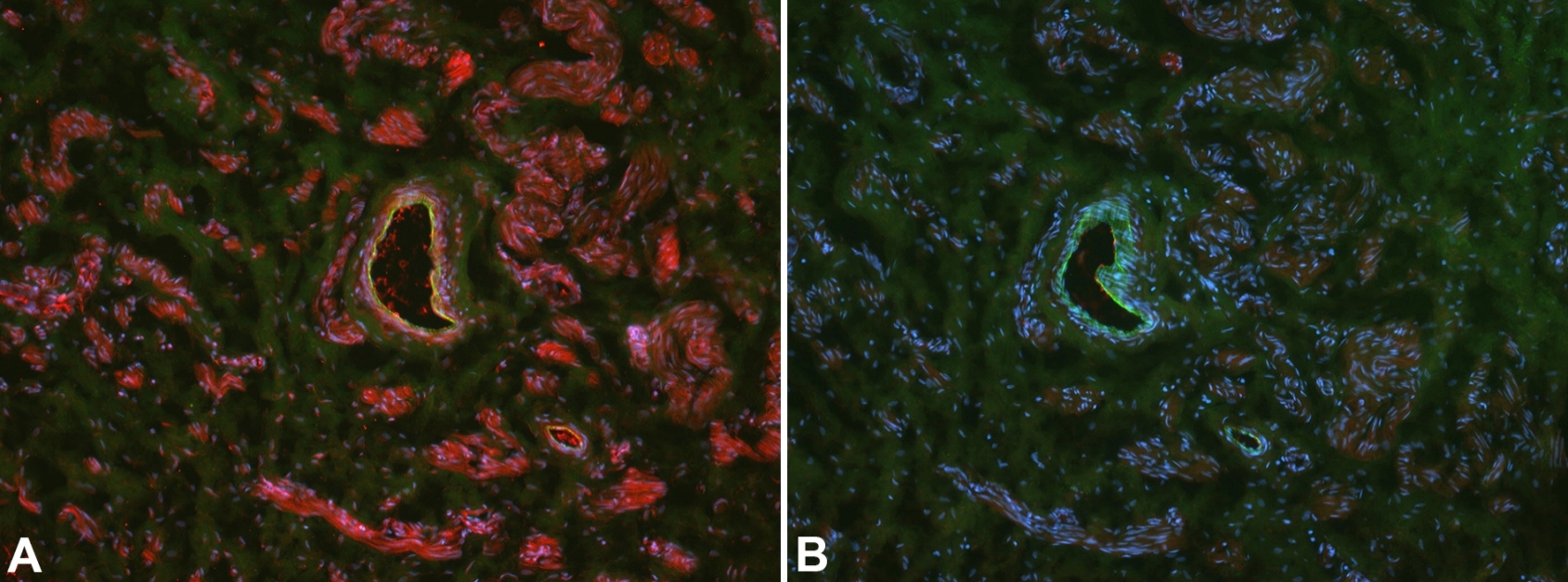
- Immunofluorescence [A] (red) with a polyclonal rabbit anti-human ccbe1 (K27876) antibody on human foreskin. The section was fixed with 4% PFA for 25 min, the antibody was diluted 1:100. [B] Control without primary antibody (yellow in A and green in B correponds to the autofluorescence within the Membrana elastica interna of an artery). A signal is visible in fibrocytes, smooth muscle cells and probably in endothelial cells. The experiment was performed by the research group of Prof. Dr. J. Wilting, University Göttingen, Germany.
- Submitted by
- ReliaTech GmbH (provider)
- Main image

- Experimental details
- Immunofluorescence [A] (red) with a polyclonal rabbit anti-human ccbe1 (K27875) antibody on human placenta tissue. The section was fixed with 4% PFA for 25 min, the antibody was diluted 1:100. [B] Control without primary antibody. A signal is visible in fibrocytes, smooth muscle cells and probably in endothelial cells. The experiment was performed by the research group of Prof. Dr. J. Wilting, University Göttingen, Germany.
- Sample type
- human placenta tissue

Supportive validation
- Submitted by
- ReliaTech GmbH (provider)
- Main image

- Experimental details
- Immunofluorescence staining (green) of human foreskin (cryo-section of unfixed tissue) with anti human ccbe1 (K6039; dilution 1:50) [Cat# 102-PA36]. A) Note specific staining in epidermis (ep) and in scattered cells in the dermis. B) Negative control of a consecutive section. Nuclei counter-stained with Dapi (blue). Specimen provided by Prof. Dr. J. Wilting, Goettingen. The experiment was performed by the research group of Prof. Dr. J. Wilting, University Göttingen, Germany.
- Sample type
- Human Foreskin

- Submitted by
- ReliaTech GmbH (provider)
- Main image
- Experimental details
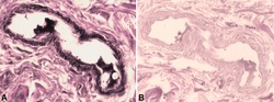
- Immunoperoxidase staining with a polyclonal rabbit anti-human ccbe1 (K27876) antibody on human skin. The section was fixed with 4% PFA overnight, the antibody was diluted 1:100. B) Control without primary antibody. A signal is visible in smooth muscle cells, a little bit weaker in endothelial cells as well as in the connective tissue. The experiment was performed by the research group of Prof. Dr. J. Wilting, University Göttingen, Germany.
- Sample type
- human skin
