 Explore
 Validate
 Learn
Learn Western blot
 ELISA
ELISAAntibody data
- Antibody Data
- Antigen structure
- References [2]
- Comments [0]
- Validations
- Western blot [2]
- Immunohistochemistry [1]
- Flow cytometry [4]
Submit
Validation data
Reference
Comment
Report error
- Product number
- NBP1-49311 - Provider product page

- Provider
- Novus Biologicals
- Proper citation
- Novus Cat#NBP1-49311, RRID:AB_10011458
- Product name
- Mouse Monoclonal CLEC4E Antibody
- Antibody type
- Monoclonal
- Description
- Protein G purified.
- Reactivity
- Human, Rat
- Host
- Mouse
- Isotype
- IgG
- Vial size
- 0.1 ml
- Concentration
- 1 mg/ml
- Storage
- Store at 4C short term. Aliquot and store at -20C long term. Avoid freeze-thaw cycles.
Submitted references C-type lectin receptors Mcl and Mincle control development of multiple sclerosis-like neuroinflammation.
Mincle signaling in the innate immune response after traumatic brain injury.
N'diaye M, Brauner S, Flytzani S, Kular L, Warnecke A, Adzemovic MZ, Piket E, Min JH, Edwards W, Mela F, Choi HY, Magg V, James T, Linden M, Reichardt HM, Daws MR, van Horssen J, Kockum I, Harris RA, Olsson T, Guerreiro-Cacais AO, Jagodic M
The Journal of clinical investigation 2020 Feb 3;130(2):838-852
The Journal of clinical investigation 2020 Feb 3;130(2):838-852
Mincle signaling in the innate immune response after traumatic brain injury.
de Rivero Vaccari JC, Brand FJ 3rd, Berti AF, Alonso OF, Bullock MR, de Rivero Vaccari JP
Journal of neurotrauma 2015 Feb 15;32(4):228-36
Journal of neurotrauma 2015 Feb 15;32(4):228-36
No comments: Submit comment
Supportive validation
- Submitted by
- Novus Biologicals (provider)
- Main image

- Experimental details
- Western Blot: CLEC4E Antibody (16E3) [NBP1-49311] - Cell lysates of HeLa (35ug) were resolved by SDS-PAGE, transferred to NC membrane and probed with anti-human CLEC4E (1:1000). Proteins were visualized using a goat anti-mouse secondary antibody conjugated to HRP and an ECL detection system.

- Submitted by
- Novus Biologicals (provider)
- Main image

- Experimental details
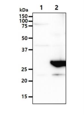
- Western Blot: CLEC4E Antibody (16E3) [NBP1-49311] - The Cell lysates (40ug) were resolved by SDS-PAGE, transferred to PVDF membrane and probed with anti-human MINCLE antibody (1:1000). Proteins were visualized using a goat anti-mouse secondary antibody conjugated to HRP and an ECL detection system. Lane 1. : 293T cell lysate Lane 2. : CLEC4E transfected 293T cell lysate
Supportive validation
- Submitted by
- Novus Biologicals (provider)
- Main image

- Experimental details
- Immunohistochemistry-Paraffin: CLEC4E Antibody (16E3) [NBP1-49311] - Paraffin embedded sections of colorectal cancer tissue were incubated with anti-human MINCLE antibody (1:50) for 2 hours at room temperature. Antigen retrieval was performed in 0.1M sodium citrate buffer and detected using Diaminobenzidine (DAB).

Supportive validation
- Submitted by
- Novus Biologicals (provider)
- Main image

- Experimental details
- Flow Cytometry: CLEC4E Antibody (16E3) [NBP1-49311] - Flow cytometry analysis of CLEC4E in LNCap cell line, staining at 2-5ug for 1x10⁶cells (red line). The secondary antibody used goat anti-mouse IgG Alexa fluor 488 conjugate.Isotype control antibody was mouse IgG (black line).

- Submitted by
- Novus Biologicals (provider)
- Main image

- Experimental details
- Flow Cytometry: CLEC4E Antibody (16E3) [NBP1-49311] - An intracellular stain was performed on U-937 cells with CLEC4E Antibody (16E3) NBP1-49311AF488 (blue) and a matched isotype control (orange). Cells were fixed with 4% PFA and then permeablized with 0.1% saponin. Cells were incubated in an antibody dilution of 5 ug/mL for 30 minutes at room temperature. Both antibodies were conjugated to Alexa Fluor 488.

- Submitted by
- Novus Biologicals (provider)
- Main image
- Experimental details
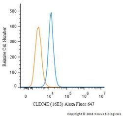
- Flow Cytometry: CLEC4E Antibody (16E3) [NBP1-49311] - An intracellular stain was performed on U87MG cells with CLEC4E Antibody (16E3) NBP1-49311AF647 (blue) and a matched isotype control (orange). Cells were fixed with 4% PFA and then permeablized with 0.1% saponin. Cells were incubated in an antibody dilution of 2.5 ug/mL for 30 minutes at room temperature. Both antibodies were conjugated to Alexa Fluor 647
- Submitted by
- Novus Biologicals (provider)
- Main image

- Experimental details
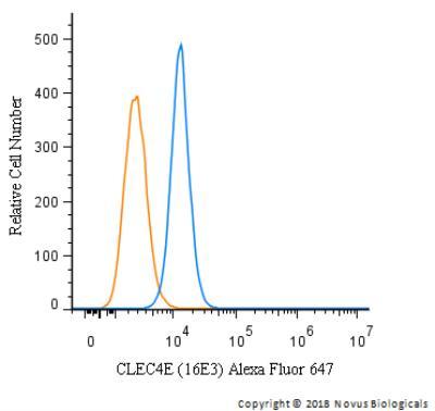
- Flow Cytometry: CLEC4E Antibody (16E3) [NBP1-49311] - An intracellular stain was performed on U87MG cells with CLEC4E Antibody (16E3) NBP1-49311AF488 (blue) and a matched isotype control (orange). Cells were fixed with 4% PFA and then permeablized with 0.1% saponin. Cells were incubated in an antibody dilution of 5 ug/mL for 30 minutes at room temperature. Both antibodies were conjugated to Alexa Fluor 488.
