Explore
 Validate
 Learn
Learn Western blot
 ELISA
ELISAAntibody data
- Antibody Data
- Antigen structure
- References [2]
- Comments [0]
- Validations
- Western blot [1]
- Immunohistochemistry [1]
- Flow cytometry [5]
Submit
Validation data
Reference
Comment
Report error
- Product number
- NBP1-51645 - Provider product page

- Provider
- Novus Biologicals
- Proper citation
- Novus Cat#NBP1-51645, RRID:AB_11022534
- Product name
- Mouse Monoclonal Rictor Antibody
- Antibody type
- Monoclonal
- Description
- Ammonium sulfate precipitation.
- Reactivity
- Human, Mouse, Simian
- Host
- Mouse
- Isotype
- IgG
- Vial size
- 0.1 ml
- Concentration
- 1.0 mg/ml
- Storage
- Store at 4C short term. Aliquot and store at -20C long term. Avoid freeze-thaw cycles.
Submitted references Loss of mTOR signaling affects cone function, cone structure and expression of cone specific proteins without affecting cone survival.
Inhibition of EGFR-AKT axis results in the suppression of ovarian tumors in vitro and in preclinical mouse model.
Ma S, Venkatesh A, Langellotto F, Le YZ, Hall MN, Rüegg MA, Punzo C
Experimental eye research 2015 Jun;135:1-13
Experimental eye research 2015 Jun;135:1-13
Inhibition of EGFR-AKT axis results in the suppression of ovarian tumors in vitro and in preclinical mouse model.
Loganathan S, Kandala PK, Gupta P, Srivastava SK
PloS one 2012;7(8):e43577
PloS one 2012;7(8):e43577
No comments: Submit comment
Supportive validation
- Submitted by
- Novus Biologicals (provider)
- Main image
- Experimental details
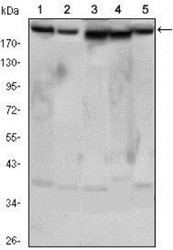
- Western Blot: Rictor Antibody (7B3) [NBP1-51645] - Western blot analysis using RICTOR mouse mAb against Hela (1), PANC-1 (2), MOLT4 (3), HepG2 (4) and HEK293 (5) cell lysates.

Supportive validation
- Submitted by
- Novus Biologicals (provider)
- Main image
- Experimental details
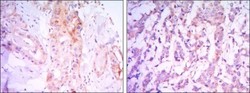
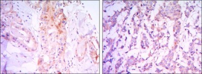
- Immunohistochemistry-Paraffin: Rictor Antibody (7B3) [NBP1-51645] - Immunohistochemical analysis of paraffin-embedded thyroid gland tissues (left) and human breast carcinoma (right) using RICTOR mouse mAb with DAB staining.
Supportive validation
- Submitted by
- Novus Biologicals (provider)
- Main image
- Experimental details
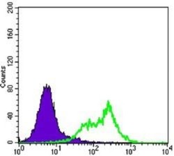
- Flow Cytometry: Rictor Antibody (7B3) [NBP1-51645] - Flow cytometric analysis of Hela cells using RICTOR mouse mAb (green) and negative control (purple).
- Submitted by
- Novus Biologicals (provider)
- Main image
- Experimental details
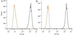
- Flow Cytometry: Rictor Antibody (7B3) [NBP1-51645] - Intracellular flow cytometric staining of 1 x 10^6 CHO (A) and HEK-293 (B) cells using RICTOR antibody (dark blue). Isotype control shown in orange. An antibody concentration of 1 ug/1x10^6 cells was used.
- Submitted by
- Novus Biologicals (provider)
- Main image
- Experimental details
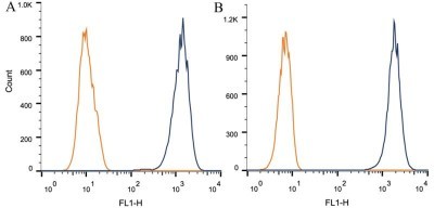
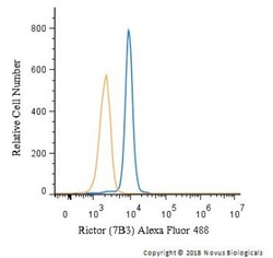
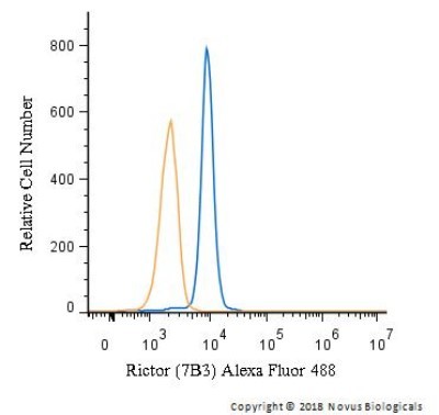
- Flow Cytometry: Rictor Antibody (7B3) [NBP1-51645] - An intracellular stain was performed on Jurkat cells with Rictor Antibody (7B3) NBP1-51645AF488 (blue) and a matched isotype control (orange). Cells were fixed with 4% PFA and then permeabilized with 0.1% saponin. Cells were incubated in an antibody dilution of 5 ug/mL for 30 minutes at room temperature. Both antibodies were conjugated to Alexa Fluor 488.
- Submitted by
- Novus Biologicals (provider)
- Main image
- Experimental details
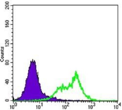
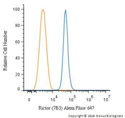
- Flow Cytometry: Rictor Antibody (7B3) [NBP1-51645] - An intracellular stain was performed on HeLa cells with Rictor Antibody (7B3) NBP1-51645AF647 (blue) and a matched isotype control (orange). Cells were fixed with 4% PFA and then permeabilized with 0.1% saponin. Cells were incubated in an antibody dilution of 2.5 ug/mL for 30 minutes at room temperature. Both antibodies were conjugated to Alexa Fluor 647.

- Submitted by
- Novus Biologicals (provider)
- Main image

- Experimental details
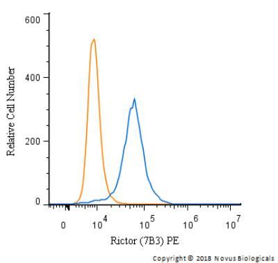
- Flow Cytometry: Rictor Antibody (7B3) [NBP1-51645] - An intracellular stain was performed on HeLa cells with Rictor Antibody (7B3) NBP1-51645PE (blue) and a matched isotype control (orange). Cells were fixed with 4% PFA and then permeabilized with 0.1% saponin. Cells were incubated in an antibody dilution of 2.5 ug/mL for 30 minutes at room temperature. Both antibodies were conjugated to Phycoerythrin.