 Explore
 Validate
 Learn
Learn Western blot
 Immunocytochemistry
Antibody data
- Antibody Data
- Antigen structure
- References [2]
- Comments [0]
- Validations
- Western blot [3]
- Immunocytochemistry [1]
Submit
Validation data
Reference
Comment
Report error
- Product number
- GTX631209 - Provider product page

- Provider
- GeneTex
- Product name
- FIS1 antibody [GT9810]
- Antibody type
- Monoclonal
- Reactivity
- Human, Mouse
- Host
- Mouse
Submitted references SIRT3 Activation by Dihydromyricetin Suppresses Chondrocytes Degeneration via Maintaining Mitochondrial Homeostasis.
Small molecule natural compound agonist of SIRT3 as a therapeutic target for the treatment of intervertebral disc degeneration.
Wang J, Wang K, Huang C, Lin D, Zhou Y, Wu Y, Tian N, Fan P, Pan X, Xu D, Hu J, Zhou Y, Wang X, Zhang X
International journal of biological sciences 2018;14(13):1873-1882
International journal of biological sciences 2018;14(13):1873-1882
Small molecule natural compound agonist of SIRT3 as a therapeutic target for the treatment of intervertebral disc degeneration.
Wang J, Nisar M, Huang C, Pan X, Lin D, Zheng G, Jin H, Chen D, Tian N, Huang Q, Duan Y, Yan Y, Wang K, Wu C, Hu J, Zhang X, Wang X
Experimental & molecular medicine 2018 Nov 12;50(11):146
Experimental & molecular medicine 2018 Nov 12;50(11):146
No comments: Submit comment
Enhanced validation
Supportive validation
- Submitted by
- GeneTex (provider)
- Enhanced method
- Genetic validation
- Main image
- Experimental details
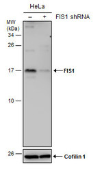
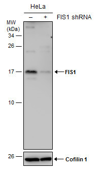
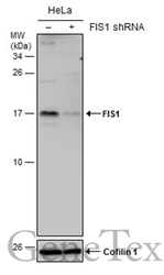
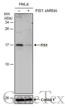
- Non-transfected (¡V) and transfected (+) HeLa whole cell extracts (30 ?g) were separated by 15% SDS-PAGE, and the membrane was blotted with FIS1 antibody [GT9810] (GTX631209) diluted at 1:500.
Supportive validation
- Submitted by
- GeneTex (provider)
- Main image
- Experimental details
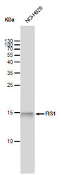
- FIS1 antibody [GT9810] detects FIS1 protein by western blot analysis. Whole cell extracts (30 £gg) was separated by 15 % SDS-PAGE, and blotted with FIS1 antibody [GT9810] (GTX631209) diluted by 1:1000

- Submitted by
- GeneTex (provider)
- Main image
- Experimental details
- Non-transfected (¡V) and transfected (+) HeLa whole cell extracts (30 ?g) were separated by 15% SDS-PAGE, and the membrane was blotted with FIS1 antibody [GT9810] (GTX631209) diluted at 1:500.
Supportive validation
- Submitted by
- GeneTex (provider)
- Main image
- Experimental details
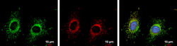
- FIS1 antibody detects FIS1 protein at mitochondria by immunofluorescent analysis.Sample: HeLa cells were fixed in 2% paraformaldehyde/culture medium at 37oC for 30 min.Green: FIS1 protein stained by FIS1 antibody (GTX631209) diluted at 1:20000.Red: MitoTrackerR Red CMXRos, a mitochondria tracker.Blue: Hoechst 33342 staining.Scale bar = 10 £gm.
