Explore
 Validate
 Learn
Learn Western blot
Antibody data
- Antibody Data
- Antigen structure
- References [0]
- Comments [0]
- Validations
- Western blot [4]
- Immunocytochemistry [2]
- Immunohistochemistry [4]
Submit
Validation data
Reference
Comment
Report error
- Product number
- PA5-53605 - Provider product page

- Provider
- Invitrogen Antibodies
- Product name
- FIS1 Polyclonal Antibody
- Antibody type
- Polyclonal
- Antigen
- Recombinant full-length protein
- Description
- Immunogen sequence: MEAVLNELVS VEDLLKFEKK FQSEKAAGSV SKSTQFEYAW CLVRSKYNDD IRKGIVLLEE LLPKGSKEEQ RDYVFYLAVG NYRLKEYEKA LKYVRGLLQT EPQNNQAKEL ERLIDKAMKK D
- Concentration
- 0.2 mg/mL
No comments: Submit comment
Supportive validation
- Submitted by
- Invitrogen Antibodies (provider)
- Main image

- Experimental details
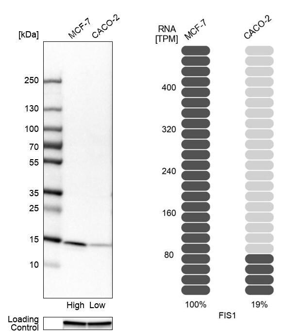
- Western blot analysis of FIS1 in human cell lines MCF-7 and Caco-2 using a FIS1 Polyclonal Antibody (Product # PA5-53605). Corresponding FIS1 RNA-seq data are presented for the same cell lines. Loading control: Anti-GAPDH.
- Submitted by
- Invitrogen Antibodies (provider)
- Main image

- Experimental details
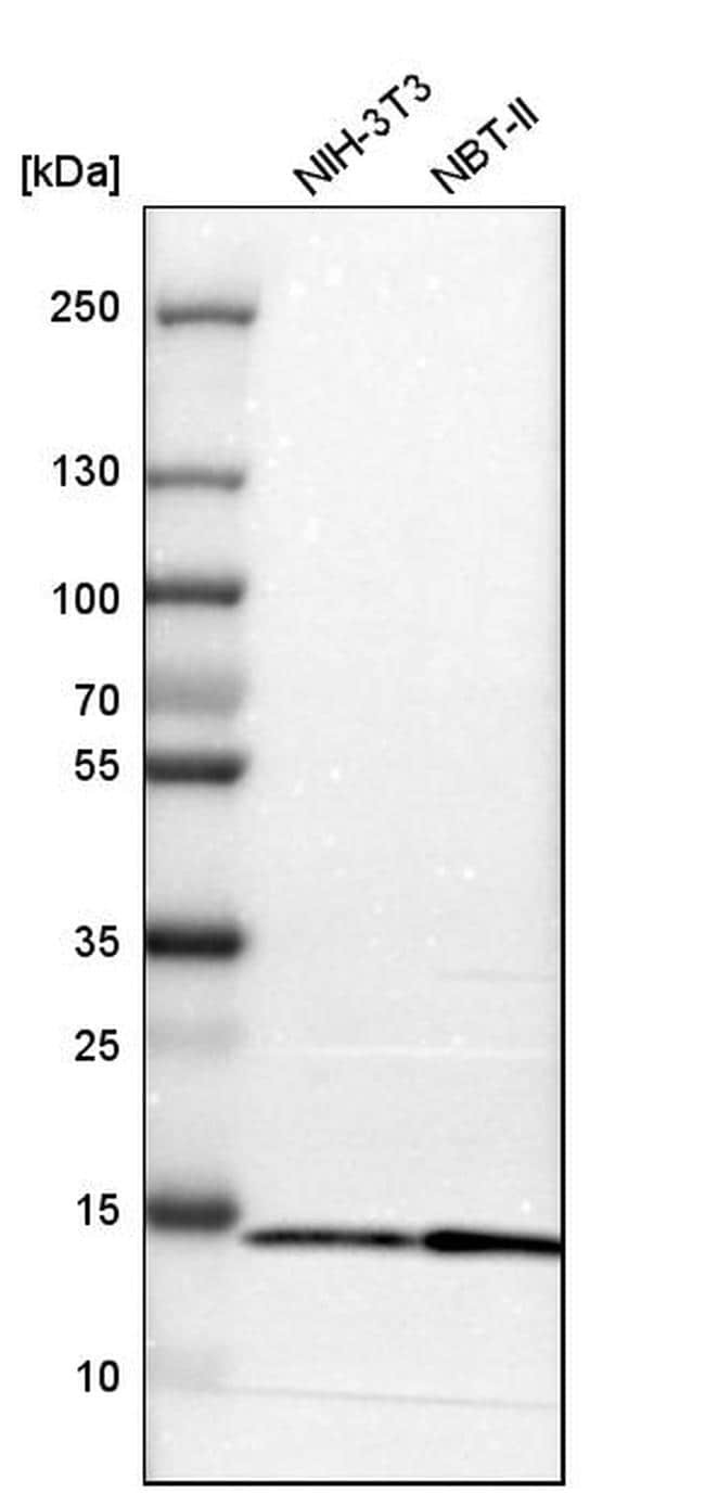
- Western blot analysis of FIS1 in mouse cell line NIH-3T3 and rat cell line NBT-II using a FIS1 Polyclonal Antibody (Product # PA5-53605).
- Submitted by
- Invitrogen Antibodies (provider)
- Main image
- Experimental details
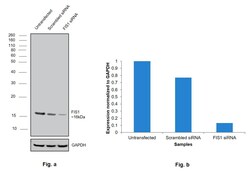
- Knockdown of FIS1 was achieved by transfecting MCF7 with FIS1 specific siRNAs (Silencer® select Product # s27265, s195204). Western Blot analysis (Fig. a) was performed using Membrane enriched extracts from the FIS1 knockdown cells (lane 3), non-targeting scrambled siRNA transfected cells (lane 2) and untransfected cells (lane 1). The blot was probed with FIS1 Polyclonal Antibody (Product # PA5-53605, 0.02 ) and Goat anti-Rabbit IgG (H+L) Superclonal™ Recombinant Secondary Antibody, HRP (Product # A27036, 1:4000). Densitometric analysis of this Western Blot is shown in histogram (Fig. b). Decrease in signal upon siRNA mediated knock down confirms that antibody is specific to FIS1.

- Submitted by
- Invitrogen Antibodies (provider)
- Main image

- Experimental details
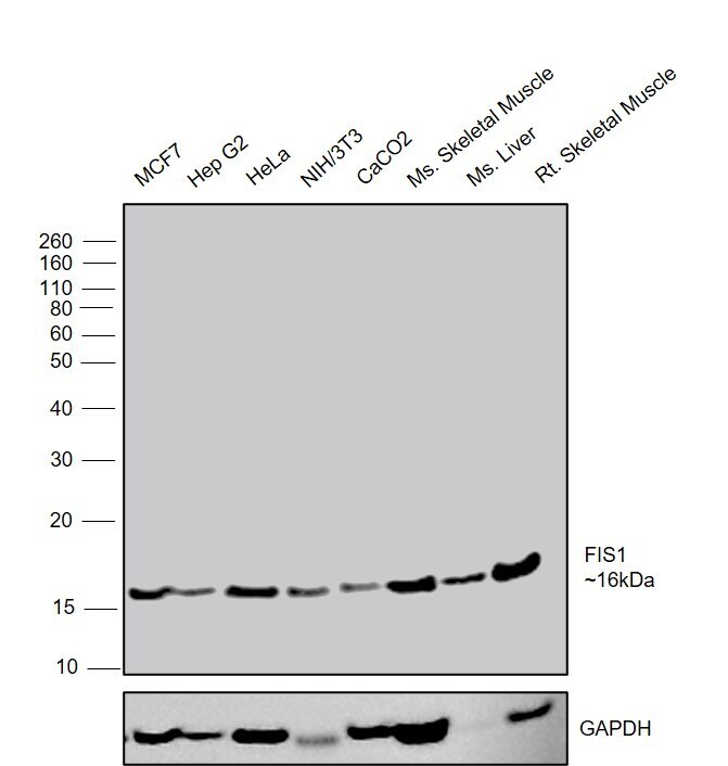
- Western Blot was performed using Anti-FIS1 Polyclonal Antibody (Product # PA5-53605) and a 16 kDa band corresponding to FIS1 was observed across cell lines and tissues tested . Membrane enriched extracts (30 µg lysate) of MCF7 (Lane 1), Hep G2 (Lane 2), HeLa (Lane 3), NIH/3T3 (Lane 4), Caco-2 (Lane 5), Mouse Skeletal Muscle (Lane 6), Mouse Liver (Lane 7), Rat Skeletal Muscle (Lane 8) were electrophoresed using NuPAGE™ 12% Bis-Tris Protein Gel (Product # NP0341BOX). Resolved proteins were then transferred onto a nitrocellulose membrane (Product # LC2001) by iBlot® 2 Dry Blotting System (Product # IB21001). The blot was probed with the primary antibody (0.04) and detected by chemiluminescence with Goat anti-Rabbit IgG (H+L) Superclonal™ Recombinant Secondary Antibody, HRP (Product # A27036, 1:4000) using the iBright FL 1000 (Product # A32752). Chemiluminescent detection was performed using Novex® ECL Chemiluminescent Substrate Reagent Kit (Product # WP20005).
Supportive validation
- Submitted by
- Invitrogen Antibodies (provider)
- Main image
- Experimental details
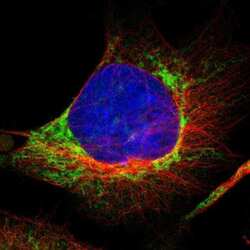
- Immunofluorescent staining of FIS1 in human cell line U-251 MG shows positivity in mitochondria. Samples were probed using a FIS1 Polyclonal Antibody (Product # PA5-53605).

- Submitted by
- Invitrogen Antibodies (provider)
- Main image
- Experimental details
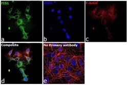
- Immunofluorescence analysis of FIS1 was performed using 70% confluent log phase Hep G2 cells. The cells were fixed with 4% paraformaldehyde for 10 minutes, permeabilized with 0.1% Triton™ X-100 for 15 minutes, and blocked with 2% BSA for 45 minutes at room temperature. The cells were labeled with FIS1 Polyclonal Antibody (Product # PA5-53605) at 1:100 in 0.1% BSA, incubated at 4 degree celsius overnight and then labeled with Donkey anti-Rabbit IgG (H+L) Highly Cross-Adsorbed Secondary Antibody, Alexa Fluor Plus 488 (Product # A32790), (1:2000), for 45 minutes at room temperature (Panel a: Green). Nuclei (Panel b:Blue) were stained with ProLong™ Diamond Antifade Mountant with DAPI (Product # P36962). F-actin (Panel c: Red) was stained with Rhodamine Phalloidin (Product # R415, 1:300). Panel d represents the merged image showing mitochondrial localization. Panel e represents control cells with no primary antibody to assess background. The images were captured at 60X magnification.

Supportive validation
- Submitted by
- Invitrogen Antibodies (provider)
- Main image
- Experimental details
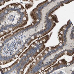
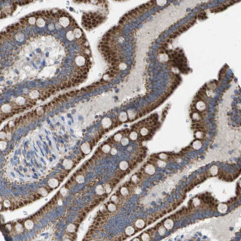
- Immunohistochemical staining of FIS1 in human small intestine using FIS1 Polyclonal Antibody (Product # PA5-53605) shows moderate granular cytoplasmic positivity in glandular cells.
- Submitted by
- Invitrogen Antibodies (provider)
- Main image

- Experimental details
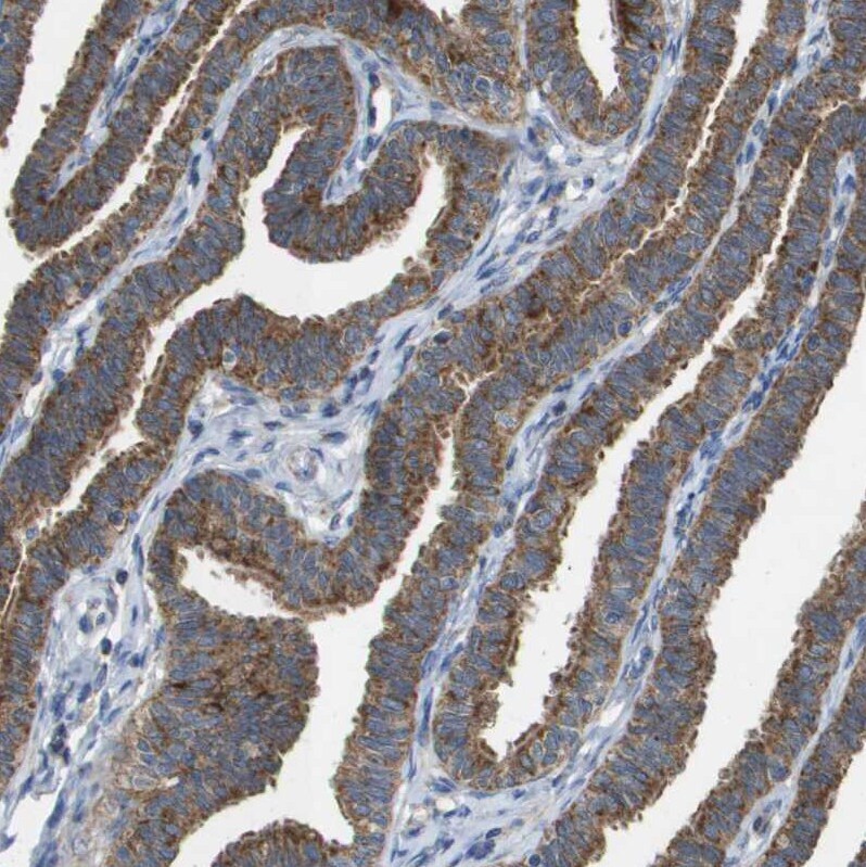
- Immunohistochemical staining of FIS1 in human Fallopian tube using FIS1 Polyclonal Antibody (Product # PA5-53605) shows moderate granular cytoplasmic positivity in glandular cells.
- Submitted by
- Invitrogen Antibodies (provider)
- Main image
- Experimental details
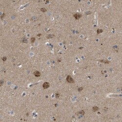
- Immunohistochemical staining of FIS1 in human cerebral cortex using FIS1 Polyclonal Antibody (Product # PA5-53605) shows moderate granular cytoplasmic positivity in neurons.

- Submitted by
- Invitrogen Antibodies (provider)
- Main image

- Experimental details
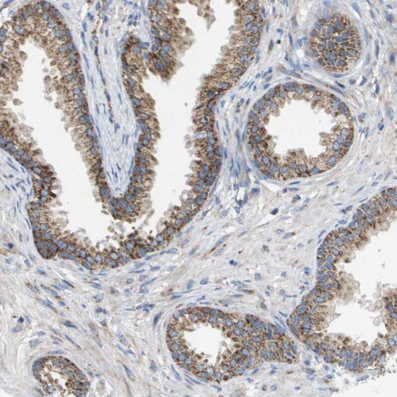
- Immunohistochemical staining of FIS1 in human prostate using FIS1 Polyclonal Antibody (Product # PA5-53605) shows moderate granular cytoplasmic positivity in glandular cells.