Explore
 Validate
 Learn
Learn Western blot
 ELISA
ELISAAntibody data
- Antibody Data
- Antigen structure
- References [3]
- Comments [0]
- Validations
- Western blot [1]
- Immunocytochemistry [1]
- Immunohistochemistry [1]
Submit
Validation data
Reference
Comment
Report error
- Product number
- 12488-1-AP - Provider product page

- Provider
- Proteintech Group
- Proper citation
- Proteintech Cat#12488-1-AP, RRID:AB_2199923
- Product name
- TBL2 antibody
- Antibody type
- Polyclonal
- Description
- KD/KO validated TBL2 antibody (Cat. #12488-1-AP) is a rabbit polyclonal antibody that shows reactivity with human, mouse, rat and has been validated for the following applications: IF, IHC, IP, WB,ELISA.
- Reactivity
- Human, Mouse, Rat
- Host
- Rabbit
- Conjugate
- Unconjugated
- Isotype
- IgG
- Vial size
- 20ul, 150ul
Submitted references TBL2 Associates With ATF4 mRNA Via Its WD40 Domain and Regulates Its Translation During ER Stress.
The endoplasmic reticulum-localized protein TBL2 interacts with the 60S ribosomal subunit.
TBL2 is a novel PERK-binding protein that modulates stress-signaling and cell survival during endoplasmic reticulum stress.
Tsukumo Y, Tsukahara S, Furuno A, Iemura S, Natsume T, Tomida A
Journal of cellular biochemistry 2016 Feb;117(2):500-9
Journal of cellular biochemistry 2016 Feb;117(2):500-9
The endoplasmic reticulum-localized protein TBL2 interacts with the 60S ribosomal subunit.
Tsukumo Y, Tsukahara S, Furuno A, Iemura S, Natsume T, Tomida A
Biochemical and biophysical research communications 2015 Jul 10;462(4):383-8
Biochemical and biophysical research communications 2015 Jul 10;462(4):383-8
TBL2 is a novel PERK-binding protein that modulates stress-signaling and cell survival during endoplasmic reticulum stress.
Tsukumo Y, Tsukahara S, Furuno A, Iemura S, Natsume T, Tomida A
PloS one 2014;9(11):e112761
PloS one 2014;9(11):e112761
No comments: Submit comment
Supportive validation
- Submitted by
- Proteintech Group (provider)
- Main image

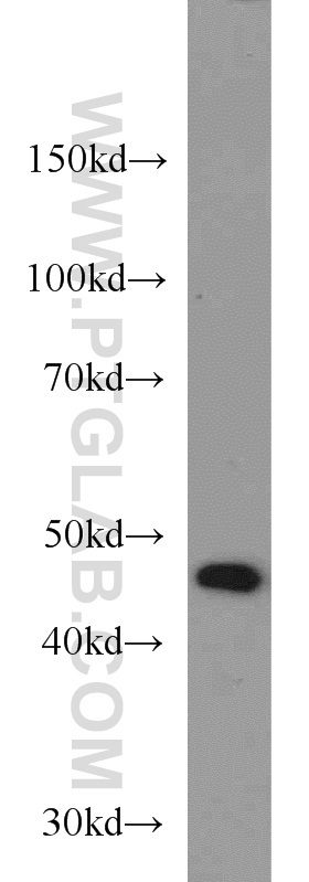
- Experimental details
- COLO 320 cells were subjected to SDS PAGE followed by western blot with 12488-1-AP(TBL2 antibody) at dilution of 1:500
- Sample type
- cell line
Supportive validation
- Submitted by
- Proteintech Group (provider)
- Main image

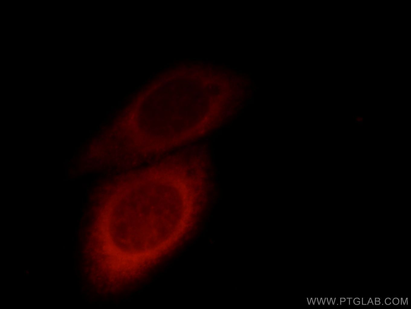
- Experimental details
- Immunofluorescent analysis of HepG2 cells, using TBL2 antibody 11640-1-AP at 1:25 dilution and Rhodamine-labeled goat anti-rabbit IgG (red).
- Sample type
- cell line
Supportive validation
- Submitted by
- Proteintech Group (provider)
- Main image

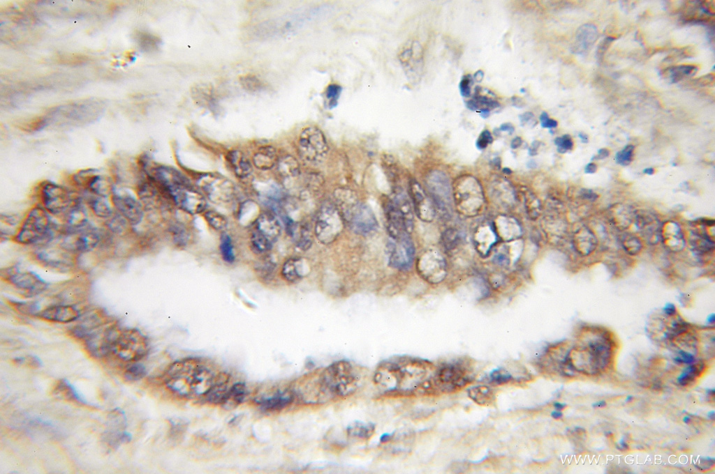
- Experimental details
- Immunohistochemical of paraffin-embedded human pancreas cancer using 12488-1-AP(TBL2 antibody) at dilution of 1:50 (under 10x lens)
- Sample type
- tissue