Explore
 Validate
 Learn
Learn Western blot
 Immunocytochemistry
Antibody data
- Antibody Data
- Antigen structure
- References [2]
- Comments [0]
- Validations
- Western blot [5]
- Immunohistochemistry [6]
Submit
Validation data
Reference
Comment
Report error
- Product number
- NBP2-20112 - Provider product page

- Provider
- Novus Biologicals
- Product name
- Rabbit Polyclonal RBPMS Antibody
- Antibody type
- Polyclonal
- Description
- Immunogen affinity purified.
- Reactivity
- Human, Mouse, Rat, Simian
- Host
- Rabbit
- Isotype
- IgG
- Vial size
- 0.1 ml
- Storage
- Aliquot and store at -20C or -80C. Avoid freeze-thaw cycles.
Submitted references Nicotinamide and WLD(S) Act Together to Prevent Neurodegeneration in Glaucoma.
GlyCAM1 negatively regulates monocyte entry into the optic nerve head and contributes to radiation-based protection in glaucoma.
Williams PA, Harder JM, Foxworth NE, Cardozo BH, Cochran KE, John SWM
Frontiers in neuroscience 2017;11:232
Frontiers in neuroscience 2017;11:232
GlyCAM1 negatively regulates monocyte entry into the optic nerve head and contributes to radiation-based protection in glaucoma.
Williams PA, Braine CE, Foxworth NE, Cochran KE, John SWM
Journal of neuroinflammation 2017 Apr 26;14(1):93
Journal of neuroinflammation 2017 Apr 26;14(1):93
No comments: Submit comment
Supportive validation
- Submitted by
- Novus Biologicals (provider)
- Main image
- Experimental details
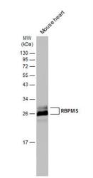
- Western Blot: RBPMS Antibody [NBP2-20112] - Mouse tissue extract (50 ug) was separated by 12% SDS-PAGE, and the membrane was blotted with RBPMS antibody diluted at 1:3000. The HRP-conjugated anti-rabbit IgG antibody (NBP2-19301) was used to detect the primary antibody.
- Submitted by
- Novus Biologicals (provider)
- Main image
- Experimental details
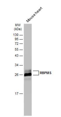
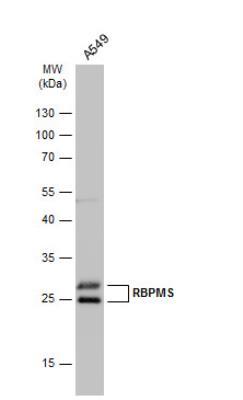
- Western Blot: RBPMS Antibody [NBP2-20112] - Whole cell extract (30 ug) was separated by 12% SDS-PAGE, and the membrane was blotted with RBPMS antibody diluted at 1:1000. The HRP-conjugated anti-rabbit IgG antibody (NBP2-19301) was used to detect the primary antibody.
- Submitted by
- Novus Biologicals (provider)
- Main image
- Experimental details
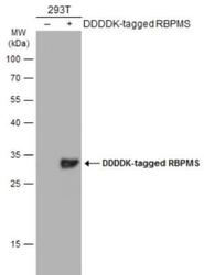
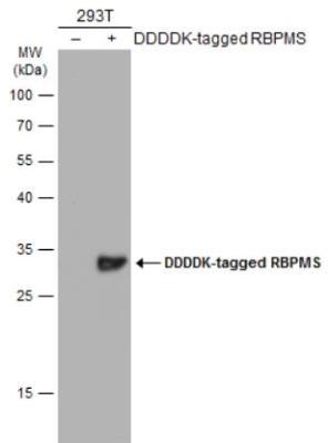
- Western Blot: RBPMS Antibody [NBP2-20112] - Non-transfected (-) and transfected (+) 293T whole cell extracts (30 ug) were separated by 12% SDS-PAGE, and the membrane was blotted with RBPMS antibody diluted at 1:1000. The HRP-conjugated anti-rabbit IgG antibody (NBP2-19301) was used to detect the primary antibody.
- Submitted by
- Novus Biologicals (provider)
- Main image
- Experimental details
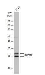
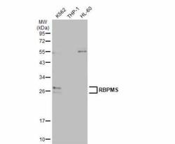
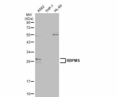
- Western Blot: RBPMS Antibody [NBP2-20112] - Various whole cell extracts (30 ug) were separated by 12% SDS-PAGE, and the membrane was blotted with RBPMS antibody diluted at 1:500. The HRP-conjugated anti-rabbit IgG antibody (NBP2-19301) was used to detect the primary antibody.
- Submitted by
- Novus Biologicals (provider)
- Main image

- Experimental details
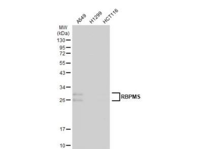
- Western Blot: RBPMS Antibody [NBP2-20112] - Various whole cell extracts (30 ug) were separated by 12% SDS-PAGE, and the membrane was blotted with RBPMS antibody diluted at 1:500. The HRP-conjugated anti-rabbit IgG antibody (NBP2-19301) was used to detect the primary antibody.
Supportive validation
- Submitted by
- Novus Biologicals (provider)
- Main image
- Experimental details
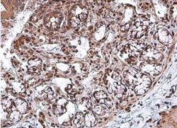
- Immunohistochemistry-Paraffin: RBPMS Antibody [NBP2-20112] - Paraffin-embedded rat colon. RBPMS antibody diluted at 1:500.

- Submitted by
- Novus Biologicals (provider)
- Main image

- Experimental details
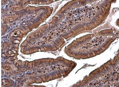
- Immunohistochemistry-Paraffin: RBPMS Antibody [NBP2-20112] - Paraffin-embedded rat duodenum. RBPMS antibody diluted at 1:500
- Submitted by
- Novus Biologicals (provider)
- Main image

- Experimental details
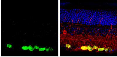
- Immunohistochemistry-Paraffin: RBPMS Antibody [NBP2-20112] - Paraffin-embedded mouse retina. Green: RBPMS protein stained by RBPMS antibody diluted at 1:250. Red: beta Tubulin 3/ Tuj1, a marker, stained by beta Tubulin 3/ Tuj1 antibody [1338] diluted at 1:500. Blue: Fluoroshield with DAPI.
- Submitted by
- Novus Biologicals (provider)
- Main image

- Experimental details
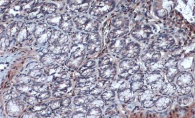
- Immunohistochemistry-Paraffin: RBPMS Antibody [NBP2-20112] - Rat colon. RBPMS stained by RBPMS antibody diluted at 1:500. Antigen Retrieval: Citrate buffer, pH 6.0, 15 min.
- Submitted by
- Novus Biologicals (provider)
- Main image

- Experimental details
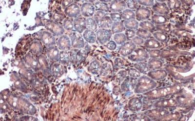
- Immunohistochemistry-Paraffin: RBPMS Antibody [NBP2-20112] - Mouse intestine. RBPMS stained by RBPMS antibody diluted at 1:2000. Antigen Retrieval: Citrate buffer, pH 6.0, 15 min.
- Submitted by
- Novus Biologicals (provider)
- Main image
- Experimental details
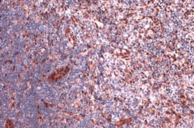
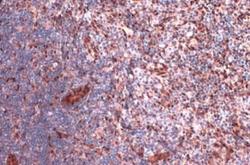
- Immunohistochemistry-Paraffin: RBPMS Antibody [NBP2-20112] - Rat lymph node. RBPMS stained by RBPMS antibody diluted at 1:2000. Antigen Retrieval: Citrate buffer, pH 6.0, 15 min.