 Explore
 Validate
 Learn
Learn Western blot
Antibody data
- Antibody Data
- Antigen structure
- References [13]
- Comments [0]
- Validations
- Western blot [4]
- Immunocytochemistry [2]
- Immunohistochemistry [4]
Submit
Validation data
Reference
Comment
Report error
- Product number
- GTX118619 - Provider product page

- Provider
- GeneTex
- Proper citation
- GeneTex Cat#GTX118619, RRID:AB_10720427
- Product name
- RBPMS antibody
- Antibody type
- Polyclonal
- Reactivity
- Human, Mouse, Rat, Simian
- Host
- Rabbit
Submitted references Evidence of Hypoxic Glial Cells in a Model of Ocular Hypertension.
RBFOX3/NeuN is dispensable for visual function.
Neuroprotective Effects of Psalmotoxin-1, an Acid-Sensing Ion Channel (ASIC) Inhibitor, in Ischemia Reperfusion in Mouse Eyes.
Neuroprotective effects of inhibitors of Acid-Sensing ion channels (ASICs) in optic nerve crush model in rodents.
Sigma-1 Receptor Regulates Mitochondrial Function in Glucose- and Oxygen-Deprived Retinal Ganglion Cells.
Damage-induced neuronal endopeptidase (DINE) enhances axonal regeneration potential of retinal ganglion cells after optic nerve injury.
Thorny ganglion cells in marmoset retina: Morphological and neurochemical characterization with antibodies against calretinin.
A mouse retinal explant model for use in studying neuroprotection in glaucoma.
Quantitative measurement of retinal ganglion cell populations via histology-based random forest classification.
C1q propagates microglial activation and neurodegeneration in the visual axis following retinal ischemia/reperfusion injury.
Endothelin-Mediated Changes in Gene Expression in Isolated Purified Rat Retinal Ganglion Cells.
Caspase-7: a critical mediator of optic nerve injury-induced retinal ganglion cell death.
Role of C/EBP homologous protein in retinal ganglion cell death after ischemia/reperfusion injury.
Jassim AH, Inman DM
Investigative ophthalmology & visual science 2019 Jan 2;60(1):1-15
Investigative ophthalmology & visual science 2019 Jan 2;60(1):1-15
RBFOX3/NeuN is dispensable for visual function.
Lin YS, Kuo KT, Chen SK, Huang HS
PloS one 2018;13(2):e0192355
PloS one 2018;13(2):e0192355
Neuroprotective Effects of Psalmotoxin-1, an Acid-Sensing Ion Channel (ASIC) Inhibitor, in Ischemia Reperfusion in Mouse Eyes.
Dibas A, Millar C, Al-Farra A, Yorio T
Current eye research 2018 Jul;43(7):921-933
Current eye research 2018 Jul;43(7):921-933
Neuroprotective effects of inhibitors of Acid-Sensing ion channels (ASICs) in optic nerve crush model in rodents.
Stankowska DL, Mueller BH 2nd, Oku H, Ikeda T, Dibas A
Current eye research 2018 Jan;43(1):84-95
Current eye research 2018 Jan;43(1):84-95
Sigma-1 Receptor Regulates Mitochondrial Function in Glucose- and Oxygen-Deprived Retinal Ganglion Cells.
Ellis DZ, Li L, Park Y, He S, Mueller B, Yorio T
Investigative ophthalmology & visual science 2017 May 1;58(5):2755-2764
Investigative ophthalmology & visual science 2017 May 1;58(5):2755-2764
Damage-induced neuronal endopeptidase (DINE) enhances axonal regeneration potential of retinal ganglion cells after optic nerve injury.
Kaneko A, Kiryu-Seo S, Matsumoto S, Kiyama H
Cell death & disease 2017 Jun 1;8(6):e2847
Cell death & disease 2017 Jun 1;8(6):e2847
Thorny ganglion cells in marmoset retina: Morphological and neurochemical characterization with antibodies against calretinin.
Chandra AJ, Lee SCS, Grünert U
The Journal of comparative neurology 2017 Dec 15;525(18):3962-3974
The Journal of comparative neurology 2017 Dec 15;525(18):3962-3974
A mouse retinal explant model for use in studying neuroprotection in glaucoma.
Pattamatta U, McPherson Z, White A
Experimental eye research 2016 Oct;151:38-44
Experimental eye research 2016 Oct;151:38-44
Quantitative measurement of retinal ganglion cell populations via histology-based random forest classification.
Hedberg-Buenz A, Christopher MA, Lewis CJ, Fernandes KA, Dutca LM, Wang K, Scheetz TE, Abràmoff MD, Libby RT, Garvin MK, Anderson MG
Experimental eye research 2016 May;146:370-385
Experimental eye research 2016 May;146:370-385
C1q propagates microglial activation and neurodegeneration in the visual axis following retinal ischemia/reperfusion injury.
Silverman SM, Kim BJ, Howell GR, Miller J, John SW, Wordinger RJ, Clark AF
Molecular neurodegeneration 2016 Mar 24;11:24
Molecular neurodegeneration 2016 Mar 24;11:24
Endothelin-Mediated Changes in Gene Expression in Isolated Purified Rat Retinal Ganglion Cells.
He S, Park YH, Yorio T, Krishnamoorthy RR
Investigative ophthalmology & visual science 2015 Sep 1;56(10):6144-61
Investigative ophthalmology & visual science 2015 Sep 1;56(10):6144-61
Caspase-7: a critical mediator of optic nerve injury-induced retinal ganglion cell death.
Choudhury S, Liu Y, Clark AF, Pang IH
Molecular neurodegeneration 2015 Aug 26;10:40
Molecular neurodegeneration 2015 Aug 26;10:40
Role of C/EBP homologous protein in retinal ganglion cell death after ischemia/reperfusion injury.
Nashine S, Liu Y, Kim BJ, Clark AF, Pang IH
Investigative ophthalmology & visual science 2014 Nov 20;56(1):221-31
Investigative ophthalmology & visual science 2014 Nov 20;56(1):221-31
No comments: Submit comment
Supportive validation
- Submitted by
- GeneTex (provider)
- Main image

- Experimental details
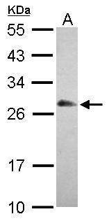
- Sample (30 ?g of whole cell lysate) A: A549 12% SDS PAGE GTX118619 diluted at 1:1000 The HRP-conjugated anti-rabbit IgG antibody (GTX213110-01) was used to detect the primary antibody.
- Submitted by
- GeneTex (provider)
- Main image
- Experimental details
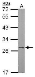
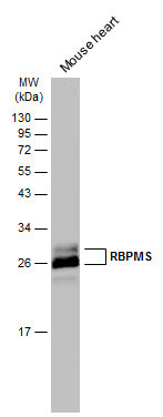
- Sample (50 ?g of whole cell lysate) A: mouse heart 12% SDS PAGE GTX118619 diluted at 1:1000 The HRP-conjugated anti-rabbit IgG antibody (GTX213110-01) was used to detect the primary antibody.

- Submitted by
- GeneTex (provider)
- Main image
- Experimental details
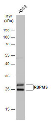
- Whole cell extract (30 ?g) was separated by 12% SDS-PAGE, and the membrane was blotted with RBPMS antibody (GTX118619) diluted at 1:1000. The HRP-conjugated anti-rabbit IgG antibody (GTX213110-01) was used to detect the primary antibody.

- Submitted by
- GeneTex (provider)
- Main image
- Experimental details
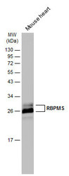
- Mouse tissue extract (50 ?g) was separated by 12% SDS-PAGE, and the membrane was blotted with RBPMS antibody (GTX118619) diluted at 1:3000. The HRP-conjugated anti-rabbit IgG antibody (GTX213110-01) was used to detect the primary antibody.
Supportive validation
- Submitted by
- GeneTex (provider)
- Main image
- Experimental details
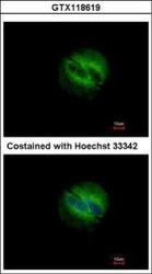
- Immunofluorescence analysis of methanol-fixed A549, using RBPMS(GTX118619) antibody at 1:500 dilution.

- Submitted by
- GeneTex (provider)
- Main image

- Experimental details
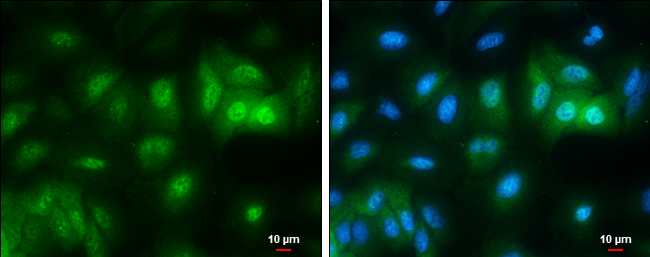
- RBPMS antibody detects RBPMS protein at cytoplasm and nucleus by immunofluorescent analysis.Sample: A549 cells were fixed in 4% paraformaldehyde at RT for 15 min.Green: RBPMS protein stained by RBPMS antibody (GTX118619) diluted at 1:500.Blue: Hoechst 33342 staining.Scale bar = 10 £gm.
Supportive validation
- Submitted by
- GeneTex (provider)
- Main image

- Experimental details
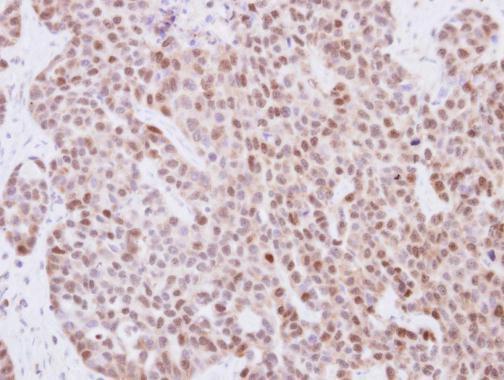
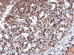
- Immunohistochemical analysis of paraffin-embedded human lung adenocarcinoma, using RBPMS(GTX118619) antibody at 1:250 dilution.
- Submitted by
- GeneTex (provider)
- Main image

- Experimental details
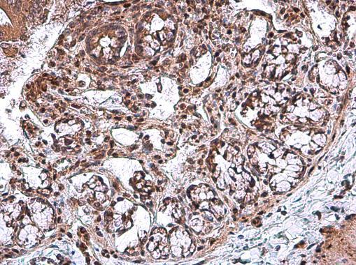
- RBPMS antibody detects RBPMS protein at cytoplasm and nucleus in rat duodenum by immunohistochemical analysis. Sample: Paraffin-embedded rat duodenum. RBPMS antibody (GTX118619) diluted at 1:500.

- Submitted by
- GeneTex (provider)
- Main image
- Experimental details
- RBPMS antibody detects RBPMS protein at cytoplasm and nucleus in rat colon by immunohistochemical analysis. Sample: Paraffin-embedded rat colon. RBPMS antibody (GTX118619) diluted at 1:500.
- Submitted by
- GeneTex (provider)
- Main image
- Experimental details
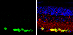
- RBPMS antibody detects RBPMS protein by immunochemical analysis.Samples: Paraffin-embedded mouse retina.Green: RBPMS protein stained by RBPMS antibody (GTX118619) diluted at 1:250.Red: beta Tubulin 3/ Tuj1, stained by beta Tubulin 3/ Tuj1 antibody [GT1338] (GTX631831) diluted at 1:500.Blue: Fluoroshield with DAPI (GTX30920).
