 Explore
 Validate
 Learn
Learn Western blot
Antibody data
- Antibody Data
- Antigen structure
- References [0]
- Comments [0]
- Validations
- Western blot [2]
- Immunocytochemistry [5]
- Immunohistochemistry [6]
Submit
Validation data
Reference
Comment
Report error
- Product number
- MA5-24641 - Provider product page

- Provider
- Invitrogen Antibodies
- Product name
- NOP56 Monoclonal Antibody (CL2603)
- Antibody type
- Monoclonal
- Antigen
- Recombinant full-length protein
- Description
- Immunogen sequence: EDPSISFSKP KKKKSFSKEE LMSSDLEETA GSTS Highest antigen sequence identity to the following orthologs: Mouse - 59%, Rat - 62%. Binds to an epitope located within the peptide sequence FSKEELMSSDLEETA as determined by overlapping synthetic peptides.
- Reactivity
- Human
- Host
- Mouse
- Isotype
- IgG
- Antibody clone number
- CL2603
- Vial size
- 100 µL
- Concentration
- 1 mg/mL
- Storage
- Store at 4°C short term. For long term storage, store at -20°C, avoiding freeze/thaw cycles.
No comments: Submit comment
Supportive validation
- Submitted by
- Invitrogen Antibodies (provider)
- Main image

- Experimental details
- Western blot analysis of NOP56 in Lane1: Marker (kDa), Lane 2:Human cell line U-251 MG. Samples were probed using a NOP56 Monoclonal Antibody (Product # MA5-24641).

- Submitted by
- Invitrogen Antibodies (provider)
- Main image

- Experimental details
- Western blot analysis of extracts from U-251 cells, transfected with: control siRNA, target specific siRNA probe #1, target specific siRNA probe #2, using NOP56 Monoclonal Antibody (CL2603) (Product # MA5-24641). Downregulation of antibody signal confirms target specificity. Remaining % intensity, relative control lane, is indicated. Anti-GAPDH monoclonal antibody was used as loading control.

Supportive validation
- Submitted by
- Invitrogen Antibodies (provider)
- Main image

- Experimental details
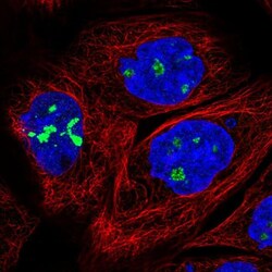
- Immunofluorescence staining of NOP56 in HeLa cell line using a NOP56 Monoclonal Antibody (Product # MA5-24641), showing nucleoli-specific staining in green. Microtubule- and nuclear probes are visualized in red and blue respectively (where available).

- Submitted by
- Invitrogen Antibodies (provider)
- Main image
- Experimental details
- Immunofluorescence staining of NOP56 in A431 cell line using a NOP56 Monoclonal Antibody (Product # MA5-24641), showing nucleoli-specific staining in green. Microtubule- and nuclear probes are visualized in red and blue respectively (where available).

- Submitted by
- Invitrogen Antibodies (provider)
- Main image

- Experimental details
- Immunofluorescence staining of NOP56 in MCF7 cell line using a NOP56 Monoclonal Antibody (Product # MA5-24641), showing nucleoli-specific staining in green. Microtubule- and nuclear probes are visualized in red and blue respectively (where available).

- Submitted by
- Invitrogen Antibodies (provider)
- Main image

- Experimental details
- Immunofluorescence staining of NOP56 in U2OS cell line using a NOP56 Monoclonal Antibody (Product # MA5-24641), showing nucleoli-specific staining in green. Microtubule- and nuclear probes are visualized in red and blue respectively (where available).

- Submitted by
- Invitrogen Antibodies (provider)
- Main image

- Experimental details
- Immunofluorescence staining of NOP56 in U251 cell line using a NOP56 Monoclonal Antibody (Product # MA5-24641), showing nucleoli-specific staining in green. Microtubule- and nuclear probes are visualized in red and blue respectively (where available).

Supportive validation
- Submitted by
- Invitrogen Antibodies (provider)
- Main image

- Experimental details
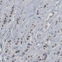
- Immunohistochemical staining of NOP56 in human prostate cancer tissue shows strong nucleolar positivity in glandular cells. Samples were probed using a NOP56 Monoclonal Antibody (Product # MA5-24641).

- Submitted by
- Invitrogen Antibodies (provider)
- Main image

- Experimental details
- Immunohistochemical staining of NOP56 in human fallopian tube shows strong nucleolar immunoreactivity in glandular and connective tissue cells. Samples were probed using a NOP56 Monoclonal Antibody (Product # MA5-24641).

- Submitted by
- Invitrogen Antibodies (provider)
- Main image
- Experimental details
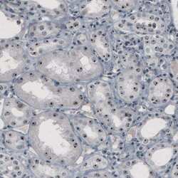
- Immunohistochemical staining of NOP56 in human kidney tissue shows strong nucleolar positivity in renal tubules. Samples were probed using a NOP56 Monoclonal Antibody (Product # MA5-24641).

- Submitted by
- Invitrogen Antibodies (provider)
- Main image

- Experimental details
- Immunohistochemical staining of NOP56 in human cervix shows strong nucleolar immunoreactivity in the epithelial cells. Samples were probed using a NOP56 Monoclonal Antibody (Product # MA5-24641).

- Submitted by
- Invitrogen Antibodies (provider)
- Main image

- Experimental details
- Immunohistochemical staining of NOP56 in human liver tissue shows strong nucleolar positivity in hepatocytes. Samples were probed using a NOP56 Monoclonal Antibody (Product # MA5-24641).

- Submitted by
- Invitrogen Antibodies (provider)
- Main image
- Experimental details
- Immunohistochemical staining of NOP56 in human breast cancer tissue shows strong nucleolar positivity in tumor cells. Samples were probed using a NOP56 Monoclonal Antibody (Product # MA5-24641).
