Explore
 Validate
 Learn
Learn Western blot
Antibody data
- Antibody Data
- Antigen structure
- References [0]
- Comments [0]
- Validations
- Western blot [3]
- Immunocytochemistry [2]
- Immunohistochemistry [3]
- Flow cytometry [1]
Submit
Validation data
Reference
Comment
Report error
- Product number
- MA5-34849 - Provider product page

- Provider
- Invitrogen Antibodies
- Product name
- NDUFAF1 Recombinant Rabbit Monoclonal Antibody (JE47-18)
- Antibody type
- Monoclonal
- Antigen
- Recombinant full-length protein
- Description
- Positive Control: K562 cell, 293T, SKOV-3, human liver cancer tissue, human prostate cancer tissue, human kidney tissue, THP-1.
- Reactivity
- Human
- Host
- Rabbit
- Isotype
- IgG
- Antibody clone number
- JE47-18
- Vial size
- 100 µL
- Concentration
- 1 mg/mL
- Storage
- -20° C, Avoid Freeze/Thaw Cycles, store in dark
No comments: Submit comment
Supportive validation
- Submitted by
- Invitrogen Antibodies (provider)
- Main image
- Experimental details
- Western blot analysis of NDUFAF1 in K562 cell lysate. Samples were transferred to PVDF membrane, blocked with 5% BSA (1 hour), incubated with NDUFAF1 monoclonal antibody (Product # MA5-34849), at a dilution of 1:1000, followed by Goat Anti-Rabbit IgG-HRP (1 hour) with a dilution of 1:5000.

- Submitted by
- Invitrogen Antibodies (provider)
- Main image
- Experimental details
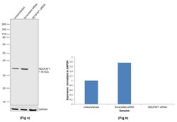
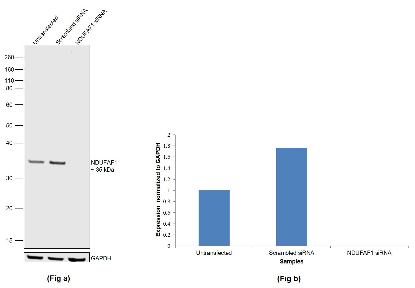
- Knockdown of Complex I intermediate-associated protein 30, mitochondrial was achieved by transfecting HEK-293 with Complex I intermediate-associated protein 30, mitochondrial specific siRNAs (Silencer® select Product # S27439, S27438). Western Blot analysis (Fig. a) was performed using Whole cell extracts from the Complex I intermediate-associated protein 30, mitochondrial knockdown cells (lane 3), non-targeting scrambled siRNA transfected cells (lane 2) and untransfected cells (lane 1). The blot was probed with NDUFAF1 Recombinant Rabbit Monoclonal Antibody (JE47-18) (Product # MA5-34849, 1:1000 ) and Goat anti-Rabbit IgG (H+L) Superclonal™ Recombinant Secondary Antibody, HRP (Product # A27036, 1:4000). Densitometric analysis of this Western Blot is shown in histogram (Fig. b). Decrease in signal upon siRNA mediated knock down confirms that antibody is specific to Complex I intermediate-associated protein 30, mitochondrial.
- Submitted by
- Invitrogen Antibodies (provider)
- Main image
- Experimental details
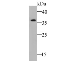
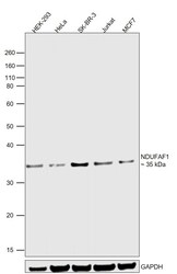
- Western Blot was performed using Anti-NDUFAF1 Recombinant Rabbit Monoclonal Antibody (JE47-18) (Product # MA5-34849) and a 35 kDa band corresponding to Complex I intermediate-associated protein 30, mitochondrial was observed. Whole cell extracts (30 µg lysate) of HEK-293 (Lane 1), HeLa (Lane 2), SK-BR-3 (Lane 3), Jurkat (Lane 4) and MCF7 (Lane 5) were electrophoresed using NuPAGE™ 10% Bis-Tris Protein Gel (Product # NP0302BOX). Resolved proteins were then transferred onto a nitrocellulose membrane (Product # IB23001) by iBlot® 2 Dry Blotting System (Product # IB21001). The blot was probed with the primary antibody (1:1000) and detected by chemiluminescence with Goat anti-Rabbit IgG (H+L) Superclonal™ Recombinant Secondary Antibody, HRP (Product # A27036, 1:4000) using the iBright FL 1000 (Product # A32752). Chemiluminescent detection was performed using Novex® ECL Chemiluminescent Substrate Reagent Kit (Product # WP20005).
Supportive validation
- Submitted by
- Invitrogen Antibodies (provider)
- Main image
- Experimental details
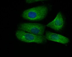
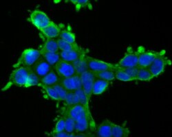
- Immunofluorescent analysis of NDUFAF1 in SKOV-3 cells (green). Samples were formalin fixed, permeabilized with 0.1% Triton X-100 in TBS (1 hour, room temperature) and blocked with 1% BSA (15 min, room temperature), incubated with NDUFAF1 monoclonal antibody (Product # MA5-34849) at a dilution of 1:200 (1 hour, room temperature), and followed by Alexa Fluor 488 Goat anti-Rabbit IgG and DAPI (blue) with a dilution of 1:100.
- Submitted by
- Invitrogen Antibodies (provider)
- Main image
- Experimental details
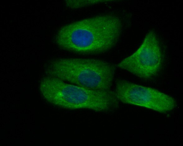
- Immunofluorescent analysis of NDUFAF1 in 293T cells (green). Samples were formalin fixed, permeabilized with 0.1% Triton X-100 in TBS (1 hour, room temperature) and blocked with 1% BSA (15 min, room temperature), incubated with NDUFAF1 monoclonal antibody (Product # MA5-34849) at a dilution of 1:200 (1 hour, room temperature), and followed by Alexa Fluor 488 Goat anti-Rabbit IgG and DAPI (blue) with a dilution of 1:100.

Supportive validation
- Submitted by
- Invitrogen Antibodies (provider)
- Main image
- Experimental details
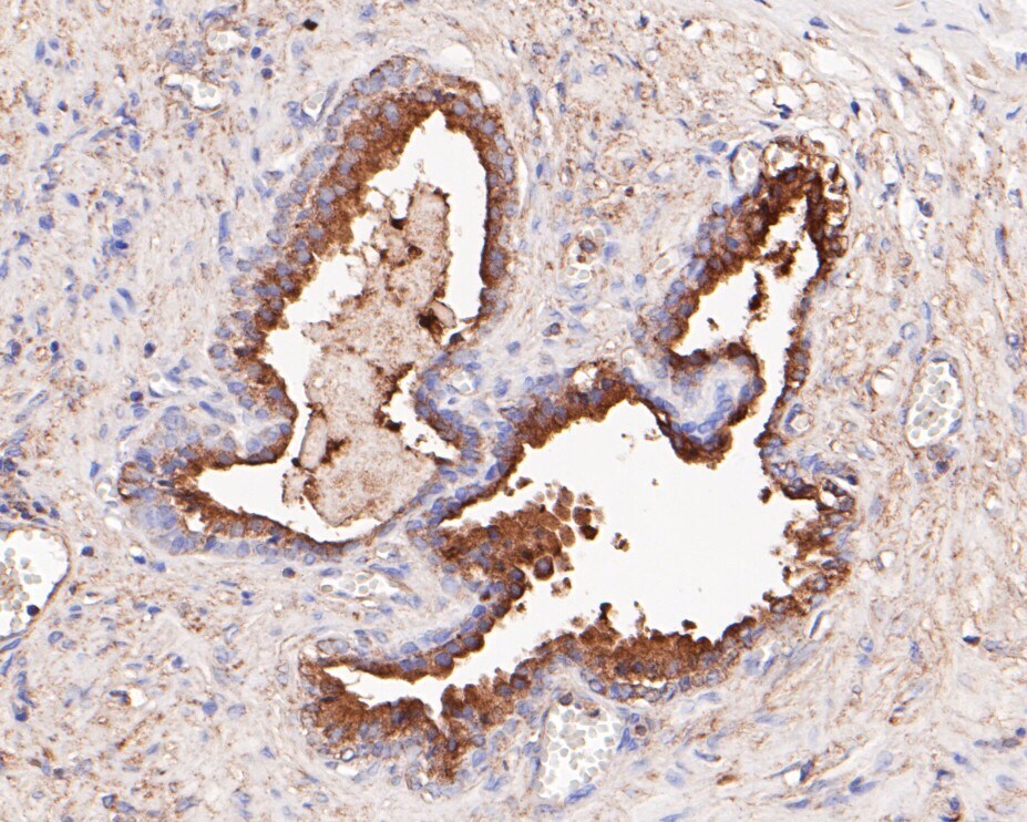
- Immunohistochemistry analysis of NDUFAF1 in paraffin-embedded human prostate cancer tissue. Samples were heat mediated antigen retrieval with Tris-EDTA buffer (pH 8.0-8.4, 20 minutes) and blocked in 5% BSA (30 min, room temperature), incubated with NDUFAF1 monoclonal antibody (Product # MA5-34849) at a dilution of 1:200 (30 min, room temperature), and followed by HRP conjugate, DAB and hematoxylin (mounted with DPX).
- Submitted by
- Invitrogen Antibodies (provider)
- Main image
- Experimental details
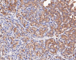
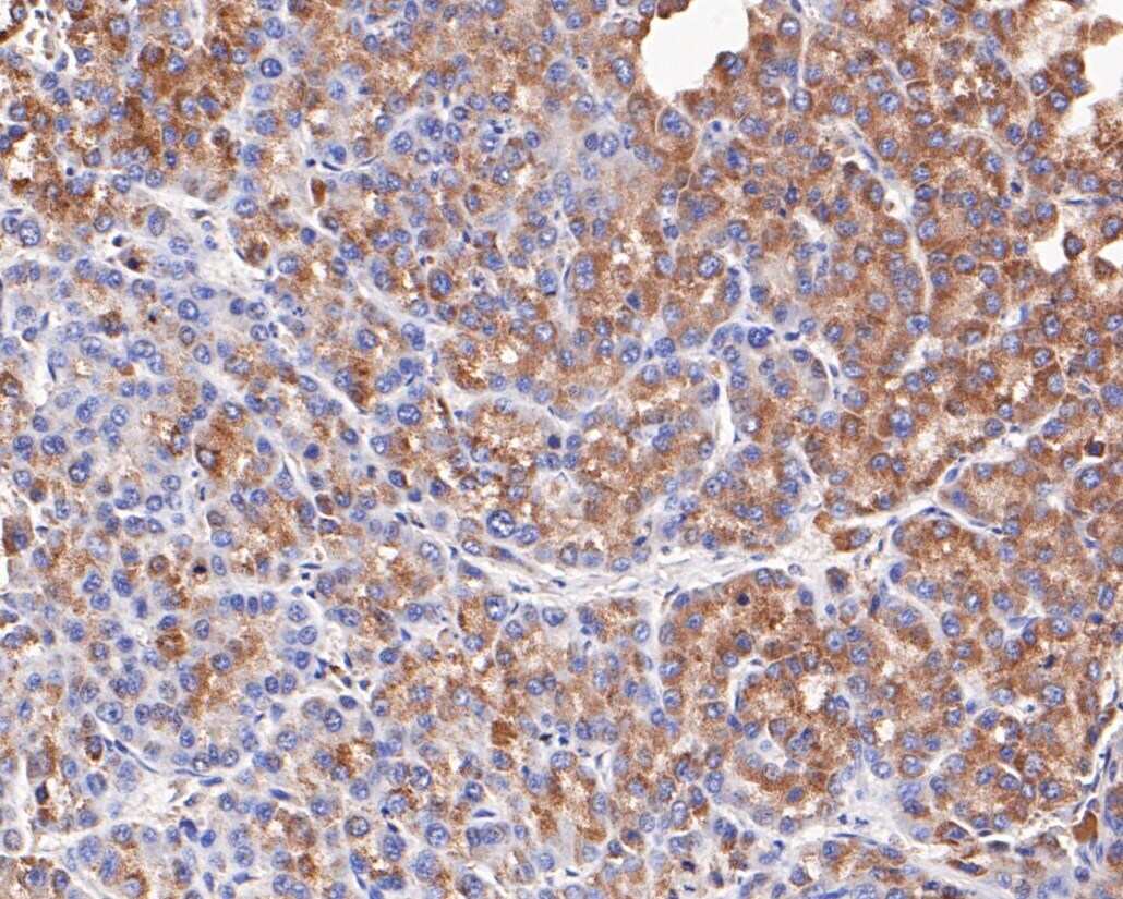
- Immunohistochemistry analysis of NDUFAF1 in paraffin-embedded human liver cancer tissue. Samples were heat mediated antigen retrieval with Tris-EDTA buffer (pH 8.0-8.4, 20 minutes) and blocked in 5% BSA (30 min, room temperature), incubated with NDUFAF1 monoclonal antibody (Product # MA5-34849) at a dilution of 1:200 (30 min, room temperature), and followed by HRP conjugate, DAB and hematoxylin (mounted with DPX).
- Submitted by
- Invitrogen Antibodies (provider)
- Main image
- Experimental details
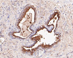
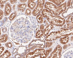
- Immunohistochemistry analysis of NDUFAF1 in paraffin-embedded human kidney tissue. Samples were heat mediated antigen retrieval with Tris-EDTA buffer (pH 8.0-8.4, 20 minutes) and blocked in 5% BSA (30 min, room temperature), incubated with NDUFAF1 monoclonal antibody (Product # MA5-34849) at a dilution of 1:200 (30 min, room temperature), and followed by HRP conjugate, DAB and hematoxylin (mounted with DPX).

Supportive validation
- Submitted by
- Invitrogen Antibodies (provider)
- Main image
- Experimental details
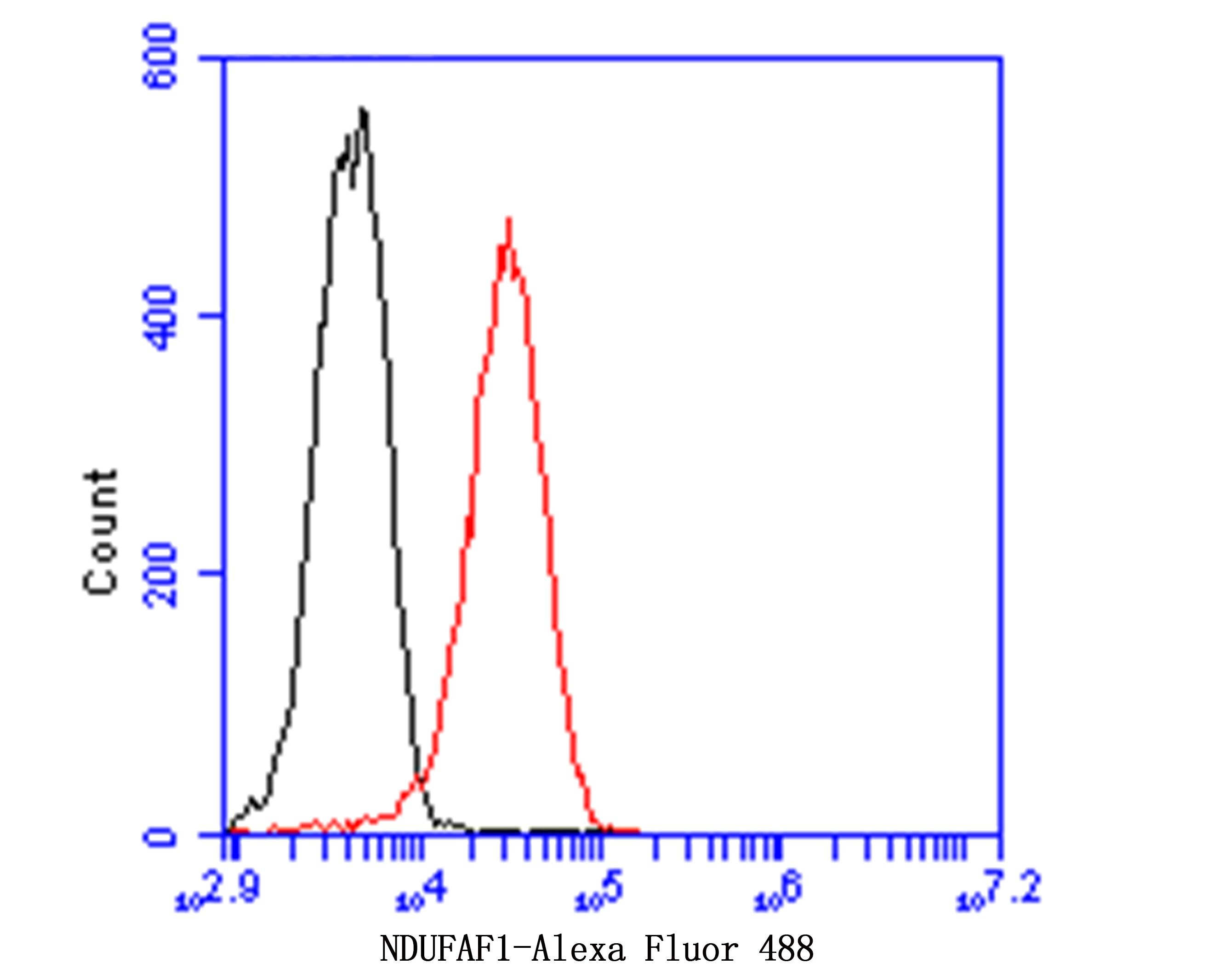
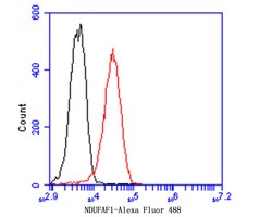
- Flow cytometry of NDUFAF1 in THP-1 cells (red) compared with an unlabelled control (cells without incubation with primary antibody; black). Samples were incubated with NDUFAF1 monoclonal antibody (Product # MA5-34849) at a dilution of 1:100, followed by Alexa Fluor 488-conjugated goat anti-rabbit IgG (30 min) with a dilution of 1:500.