 Explore
 Validate
 Learn
Learn Western blot
 Immunocytochemistry
Antibody data
- Antibody Data
- Antigen structure
- References [1]
- Comments [0]
- Validations
- Western blot [1]
- Immunocytochemistry [1]
- Immunohistochemistry [13]
Submit
Validation data
Reference
Comment
Report error
- Product number
- HPA011861 - Provider product page

- Provider
- Atlas Antibodies
- Proper citation
- Atlas Antibodies Cat#HPA011861, RRID:AB_1844493
- Product name
- Anti-ACBD5
- Antibody type
- Polyclonal
- Reactivity
- Human
- Host
- Rabbit
- Conjugate
- Unconjugated
- Antigen sequence
IQVPPGNGNIGNMQVVAVEGKGEVKHGGEDGRNNS
GAPHREKRGGETDEFSNVRRGRGHRMQHLSEGTKG
RQVGSGGDGERWGSDRGSRGSLNEQIALVLMRLQE
DMQNVLQRLQKLETLTALQAKSSTSTLQTAPQPTS
QRPSWWPF- Isotype
- IgG
- Vial size
- 100 µl
- Storage
- Store at +4°C for short term storage. Long time storage is recommended at -20°C.
Submitted references Peroxisomal Atg37 binds Atg30 or palmitoyl-CoA to regulate phagophore formation during pexophagy.
Nazarko TY, Ozeki K, Till A, Ramakrishnan G, Lotfi P, Yan M, Subramani S
The Journal of cell biology 2014 Feb 17;204(4):541-57
The Journal of cell biology 2014 Feb 17;204(4):541-57
No comments: Submit comment
Enhanced validation
- Submitted by
- Atlas Antibodies (provider)
- Enhanced method
- Orthogonal validation
- Main image
- Experimental details
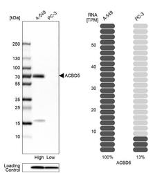
- Western blot analysis in human cell lines A-549 and PC-3 using Anti-ACBD5 antibody. Corresponding ACBD5 RNA-seq data are presented for the same cell lines. Loading control: Anti-PPIB.

Supportive validation
- Submitted by
- Atlas Antibodies (provider)
- Main image

- Experimental details
- Immunofluorescent staining of human cell line A-431 shows localization to peroxisomes.
- Sample type
- HUMAN

Enhanced validation
Enhanced validation
Supportive validation
- Submitted by
- Atlas Antibodies (provider)
- Enhanced method
- Orthogonal validation
- Main image

- Experimental details
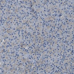
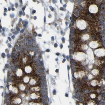
- Immunohistochemistry analysis in human small intestine and pancreas tissues using HPA011861 antibody. Corresponding ACBD5 RNA-seq data are presented for the same tissues.
- Sample type
- HUMAN

Enhanced validation
- Submitted by
- Atlas Antibodies (provider)
- Enhanced method
- Independent antibody validation
- Main image

- Experimental details
- Immunohistochemical staining of human kidney, liver, pancreas and small intestine using Anti-ACBD5 antibody HPA011861 (A) shows similar protein distribution across tissues to independent antibody HPA012145 (B).

Supportive validation
- Submitted by
- Atlas Antibodies (provider)
- Main image

- Experimental details
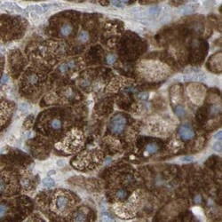
- Immunohistochemical staining of human liver shows strong cytoplasmic positivity in hepatocytes.

- Submitted by
- Atlas Antibodies (provider)
- Main image

- Experimental details
- Immunohistochemical staining of human parathyroid gland shows high expression.
- Sample type
- HUMAN

- Submitted by
- Atlas Antibodies (provider)
- Main image
- Experimental details
- Immunohistochemical staining of human pancreas shows low expression as expected.
- Sample type
- HUMAN

- Submitted by
- Atlas Antibodies (provider)
- Main image
- Experimental details
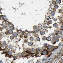
- Immunohistochemical staining of human testis using Anti-ACBD5 antibody HPA011861.
- Sample type
- HUMAN

- Submitted by
- Atlas Antibodies (provider)
- Main image

- Experimental details
- Immunohistochemical staining of human liver using Anti-ACBD5 antibody HPA011861.
- Sample type
- HUMAN

- Submitted by
- Atlas Antibodies (provider)
- Main image

- Experimental details
- Immunohistochemical staining of human colon using Anti-ACBD5 antibody HPA011861.
- Sample type
- HUMAN
- Submitted by
- Atlas Antibodies (provider)
- Main image
- Experimental details
- Immunohistochemical staining of human adrenal gland using Anti-ACBD5 antibody HPA011861.
- Sample type
- HUMAN

- Submitted by
- Atlas Antibodies (provider)
- Main image

- Experimental details
- Immunohistochemical staining of human pancreas shows very weak granular cytoplasmic positivity in exocrine glandular cells.
- Sample type
- HUMAN

- Submitted by
- Atlas Antibodies (provider)
- Main image

- Experimental details
- Immunohistochemical staining of human small intestine shows strong granular cytoplasmic positivity in glandular cells.
- Sample type
- HUMAN

- Submitted by
- Atlas Antibodies (provider)
- Main image

- Experimental details
- Immunohistochemical staining of human liver shows strong granular cytoplasmic positivity in hepatocytes.
- Sample type
- HUMAN

- Submitted by
- Atlas Antibodies (provider)
- Main image

- Experimental details
- Immunohistochemical staining of human kidney shows moderate granular cytoplasmic positivity in cells in tubules.
- Sample type
- HUMAN
