Explore
 Validate
 Learn
Learn Western blot
Antibody data
- Antibody Data
- Antigen structure
- References [0]
- Comments [0]
- Validations
- Western blot [1]
- Immunohistochemistry [1]
Submit
Validation data
Reference
Comment
Report error
- Product number
- TA319211 - Provider product page

- Provider
- OriGene
- Product name
- Rabbit polyclonal anti-NOXO1 antibody
- Antibody type
- Polyclonal
- Description
- Rabbit polyclonal anti-NOXO1 antibody
- Host
- Rabbit
- Conjugate
- Unconjugated
- Epitope
- NOXO1
- Isotype
- IgG
- Antibody clone number
- NULL
- Vial size
- 100 µg
- Concentration
- 1.17 mg/mL
No comments: Submit comment
Supportive validation
- Submitted by
- OriGene (provider)
- Main image
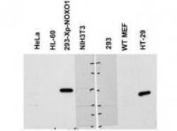
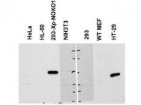
- Experimental details
- WB using Anti-NOXO1 antibody shows detection of a band ~50 kDa corresponding to human NOXO1 (arrowhead). Reactivity was observed in transfected human 293 cells and human HT-29 colon carcinoma cells (endogenous). A 1:1,000 dilution of the primary antibody was used for detection followed by secondary antibody reactivity. Specific band reactivity was competed away when the antibody was pre-incubated with the peptide immunogen (data not shown).
- Validation comment
- WB
Supportive validation
- Submitted by
- OriGene (provider)
- Main image
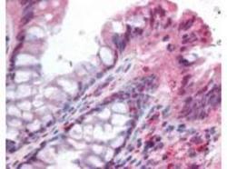
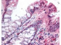
- Experimental details
- Anti-NOXO1 antibody was used at 5 ug/ml to detect signal in a variety of tissues including multi-human, multi-brain and multi-cancer slides. This image shows moderate positive staining of the lamina propia in human colon epithelium and macrophages at 40X. Tissue was formalin-fixed and paraffin embedded. The image shows localization of the antibody as the precipitated red signal, with a hematoxylin purple nuclear counterstain. Personal Communi-cation, Tina Roush, LifeSpanBiosciences, Seattle, WA.
- Validation comment
- IHC