 Explore
 Validate
 Learn
LearnNBP1-77899
antibody from Novus Biologicals
Targeting: NOXO1
P41NOXA, P41NOXB, P41NOXC, SH3PXD5, SNX28
 Western blot
 ELISA
ELISAAntibody data
- Antibody Data
- Antigen structure
- References [0]
- Comments [0]
- Validations
- Western blot [1]
- Immunohistochemistry [1]
Submit
Validation data
Reference
Comment
Report error
- Product number
- NBP1-77899 - Provider product page

- Provider
- Novus Biologicals
- Proper citation
- Novus Cat#NBP1-77899, RRID:AB_11037558
- Product name
- Rabbit Polyclonal NOXO1 Antibody
- Antibody type
- Polyclonal
- Description
- Immunogen affinity purified. A BLAST analysis was used to suggest cross reactivity with NOXO1 protein from human and chimpanzee (100% homology). Expect reactivity with alpha, delta and gamma isoforms of NOXO1. Also expect partial reactivity against NOXO1 homologues from dog and guinea pig (87%), as well as rat (75%) and mouse (68%). Reactivity against homologues from other sources is not known.
- Reactivity
- Human
- Host
- Rabbit
- Isotype
- IgG
- Vial size
- 0.1 mg
- Concentration
- 1 mg/ml
- Storage
- Store at -20C. Avoid freeze-thaw cycles.
No comments: Submit comment
Supportive validation
- Submitted by
- Novus Biologicals (provider)
- Main image

- Experimental details
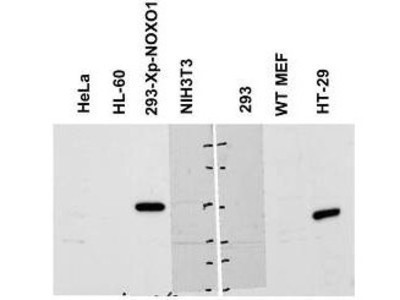
- Western Blot: NOXO1 Antibody [NBP1-77899] - NOXO1 antibody shows detection of a band ~50 kDa corresponding to human NOXO1 (arrowhead). Reactivity was observed in transfected human 293 cells and human HT-29 colon carcinoma cells (endogenous). Under these conditions endogenous NOXO1 detection was not observed in HeLa, HL-60, untransfected 293 or WT MEF cells. A 1:1,000 dilution of the primary antibody was used for detection followed by secondary antibody reactivity. Specific band reactivity was competed away when the antibody was pre-incubated with the peptide immunogen (data not shown).
Supportive validation
- Submitted by
- Novus Biologicals (provider)
- Main image
- Experimental details
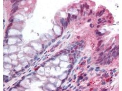
- Immunohistochemistry: NOXO1 Antibody [NBP1-77899] - Used at 5 ug/ml to detect signal in a variety of tissues including multi-human, multi-brain and multi-cancer slides. This image shows moderate positive staining of the lamina propia in human colon epithelium and macrophages at 40X. Tissue was formalin-fixed and paraffin embedded. The image shows localization of the antibody as the precipitated red signal, with a hematoxylin purple nuclear counterstain. Personal Communi-cation, Tina Roush, LifeSpanBiosciences, Seattle, WA.
