Antibody data
- Antibody Data
- Antigen structure
- References [4]
- Comments [0]
- Validations
- Immunocytochemistry [1]
- Immunohistochemistry [2]
- Other assay [4]
Submit
Validation data
Reference
Comment
Report error
- Product number
- MA1-06305 - Provider product page

- Provider
- Invitrogen Antibodies
- Product name
- K-cadherin Monoclonal Antibody (2B6)
- Antibody type
- Monoclonal
- Antigen
- Purifed from natural sources
- Description
- MA1-06305 detects K-cadherin in human and rat samples. MA1-06305 has sucessfully been used in Western blotting, immunocytochemistry, and immunohistochemistry (paraffin). By Western blotting, it detects a ~120 kDa protein representing K-cadherin. The MA1-06305 immunogen is affinity purified extracellular domain of human cadherin-6-GST fusion protein. Store at 4ºC or in small aliquots at -20ºC.
- Reactivity
- Human, Rat
- Host
- Mouse
- Isotype
- IgG
- Antibody clone number
- 2B6
- Vial size
- 100 µL
- Concentration
- 1 mg/mL
- Storage
- Store at 4°C short term. For long term storage, store at -20°C, avoiding freeze/thaw cycles.
Submitted references Angiotensin II induces coordinated calcium bursts in aldosterone-producing adrenal rosettes.
In situ molecular characterization of endoneurial microvessels that form the blood-nerve barrier in normal human adult peripheral nerves.
miR-223-3p Inhibits Human Osteosarcoma Metastasis and Progression by Directly Targeting CDH6.
Cadherin-6 is a putative tumor suppressor and target of epigenetically dysregulated miR-429 in cholangiocarcinoma.
Guagliardo NA, Klein PM, Gancayco CA, Lu A, Leng S, Makarem RR, Cho C, Rusin CG, Breault DT, Barrett PQ, Beenhakker MP
Nature communications 2020 Apr 3;11(1):1679
Nature communications 2020 Apr 3;11(1):1679
In situ molecular characterization of endoneurial microvessels that form the blood-nerve barrier in normal human adult peripheral nerves.
Ouyang X, Dong C, Ubogu EE
Journal of the peripheral nervous system : JPNS 2019 Jun;24(2):195-206
Journal of the peripheral nervous system : JPNS 2019 Jun;24(2):195-206
miR-223-3p Inhibits Human Osteosarcoma Metastasis and Progression by Directly Targeting CDH6.
Ji Q, Xu X, Song Q, Xu Y, Tai Y, Goodman SB, Bi W, Xu M, Jiao S, Maloney WJ, Wang Y
Molecular therapy : the journal of the American Society of Gene Therapy 2018 May 2;26(5):1299-1312
Molecular therapy : the journal of the American Society of Gene Therapy 2018 May 2;26(5):1299-1312
Cadherin-6 is a putative tumor suppressor and target of epigenetically dysregulated miR-429 in cholangiocarcinoma.
Goeppert B, Ernst C, Baer C, Roessler S, Renner M, Mehrabi A, Hafezi M, Pathil A, Warth A, Stenzinger A, Weichert W, Bähr M, Will R, Schirmacher P, Plass C, Weichenhan D
Epigenetics 2016 Nov;11(11):780-790
Epigenetics 2016 Nov;11(11):780-790
No comments: Submit comment
Supportive validation
- Submitted by
- Invitrogen Antibodies (provider)
- Main image
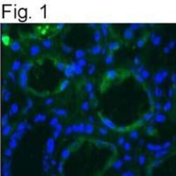
- Experimental details
- Immunostaining of K-cadherin from human skin using Product # MA1-06305.
Supportive validation
- Submitted by
- Invitrogen Antibodies (provider)
- Main image
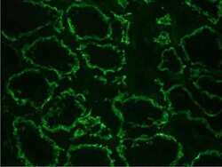
- Experimental details
- Immunohistochemistry on frozen section of human kidney: positive reactivity in renal tubules (epithelium) stained with Cadherin K monoclonal antibody (Product # MA1-06305).
- Submitted by
- Invitrogen Antibodies (provider)
- Main image
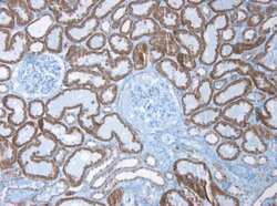
- Experimental details
- Immunohistochemistry on paraffin section of human kidney stained with Cadherin K monoclonal antibody (Product # MA1-06305).
Supportive validation
- Submitted by
- Invitrogen Antibodies (provider)
- Main image
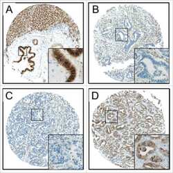
- Experimental details
- NULL
- Submitted by
- Invitrogen Antibodies (provider)
- Main image
- Experimental details
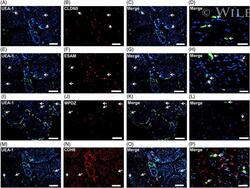
- Endothelial cell-specific proteins. Representative indirect fluorescent digital photomicrographs of cryostat axial sections of normal adult sural nerves show UEA-1-positive endothelial cells (green; A, E, I, M) with expression of CLDN5, ESAM, MPDZ, and CDH6 (red; B, F, J, N, respectively) restricted to endoneurial microvessels and fenestrated epineurial macrovessels shown in the merged images at lower and higher magnifications (yellow/ orange). CDH6 is also expressed by large diameter axons (M-P). Expression of these proteins by both endoneurial microvascular and epineurial macrovascular endothelium suggests endothelial-specific, non-restrictive barrier functions at the normal adult human blood-nerve barrier. White arrows demonstrate positively staining endoneurial microvessels. Blue (4, 6-diamidino-2-phenylindole) staining indicates nuclei. Scale bar 500 mum for A-C, E-G, I-K, M-O, and 125 mum for D, H, L and P
- Submitted by
- Invitrogen Antibodies (provider)
- Main image
- Experimental details
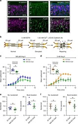
- Fig. 7 Disruption of N-cadherin shortens burst duration without affecting other calcium spike parameters. a Immunohistochemistry showing N-cadherin (red, upper left), K-cadherin (red, lower left) and F-actin (green, middle) are abundant in the zG layer (right, merged images). Scale bar: 20 um. b Model of calcium switch assay: Disrupting adherins junctional complex by reducing extracellular calcium with EDTA. Anti-N/K-cadherin antibodies (EC-Abs) binds to the now exposed extracellular N- and K-cadherin binding sites, inhibiting reformation of strong cell-cell adhesion after calcium is restored. For controls, we substituted an antibody directed to the conserved intracellular domain (IC-Ab) of cadherins. c - f Adrenal slices were stimulated with 300 pM ( c , e ) or 3 nM ( d , f ) Ang II after 1.5 min of baseline recording. EC-Abs: n = 6; IC-Ab: n = 6. c , d Mean spikes per 1 min bin over a 10 min period; EC-Abs treated slices had fewer spikes over time for both 300 pM and 3 nM Ang II (2-way ANOVA, effect of antibody: P = 0.011 and 0.018, respectively) as well as mean fractional active time ( c , d insets, mean +- SEM: 300 pM: IC-Ab 0.40 +- 0.04, EC-Abs 0.22 +- 0.03; 3 nM: IC-Ab 0.48 +- 0.05, EC-Abs 0.26 +- 0.06; Wilcoxon rank test, * P = 0.016 and 0.016, respectively). e , f Reduced activity was due to a decrease in the number of bursts per cell ( e , f left panels; mean +- SEM: 300 pM: IC-Ab 5.43 +- 0.39, EC-Abs 4.35 +- 0.44; 3 nM: IC-Ab 6.94 +- 0.67, EC-Abs 4.8 +- 0.66; Wicoxo
- Submitted by
- Invitrogen Antibodies (provider)
- Main image
- Experimental details
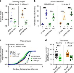
- Fig. 8 Cadherin disruption reduces both the number of clustered cells and the production of aldosterone. a Significantly fewer cells clustered after N/K EC-Abs treatment when stimulated with 300 pM or 3 nM Ang II (means +- SEM: 300 pM Ang II: IC-Ab 0.45 +- 0.19, EC-Abs 0.09 +- 0.06; 3 nM Ang II: IC-Ab 0.71 +- 0.20, EC-Abs 0.07 +- 0.04; 1-way ANOVA, 300 pM Ang II: * P = 0.026; 3 nM Ang II: *** P < 0.0001). b However, cells that remained in functional clusters were proximate (C: clustered; N-C: non-clustered; means +- SEM: 300 pM Ang II: IC-Ab-C 7.77 +- 0.50, IC-Ab-N-C 49.60 +- 4.89, EC-Abs-C 5.55 +- 1.85, EC-Abs-N-C 35.44 +- 3.78; 3 nM Ang II: IC-Ab-C 11.20 +- 1.02, IC-Ab-N-C 35.96 +- 4.13, EC-Abs-C 7.95 +- 0.58, EC-Abs-N-C 47.65 +- 6.30; 1-way ANOVA, 300pM Ang II: IC-Ab C vs C-N *** P < 0.0001, EC-Abs C vs C-N *** P < 0.0001; 3 nM Ang II: IC-Ab C vs C-N *** P = 0.0005, EC-Abs C vs C-N *** P < 0.0001) and c produced phase-locked calcium oscillations (smaller PD-STDEV). d PD-STDEV Notably, aldosterone production was reduced after N-/K-cadherin disruption, both at baseline and following 3 nM Ang II stimulation (means +- SEM: baseline: IC-Ab 1.00 +- 0.05 [ n = 23], EC-Abs 0.60 +- 0.04 [ n = 20]; 3 nM Ang II: IC-Ab 1.00 +- 0.04 [ n = 23], EC-Abs 0.70 +- 0.03 [ n = 20]; 1-way ANOVA, baseline: *** P < 0.0001; 3 nM Ang II: *** P < 0.0001). Each data point represents aldosterone secreted from slices within a well standardized to the mean production in control (IC-Ab) wells. Lines and
 Explore
 Validate
 Learn
Learn Western blot
 Immunocytochemistry