Explore
 Validate
 Learn
Learn Western blot
Antibody data
- Antibody Data
- Antigen structure
- References [1]
- Comments [0]
- Validations
- Western blot [1]
- Immunocytochemistry [1]
- Immunohistochemistry [3]
- Other assay [5]
Submit
Validation data
Reference
Comment
Report error
- Product number
- PA5-30609 - Provider product page

- Provider
- Invitrogen Antibodies
- Product name
- HEBP1 Polyclonal Antibody
- Antibody type
- Polyclonal
- Antigen
- Recombinant protein fragment
- Description
- Recommended positive controls: A549, HeLa, HepG2, HCT116.
- Concentration
- 0.63 mg/mL
Submitted references Increased expression of heme-binding protein 1 early in Alzheimer's disease is linked to neurotoxicity.
Yagensky O, Kohansal-Nodehi M, Gunaseelan S, Rabe T, Zafar S, Zerr I, Härtig W, Urlaub H, Chua JJ
eLife 2019 Aug 27;8
eLife 2019 Aug 27;8
No comments: Submit comment
Supportive validation
- Submitted by
- Invitrogen Antibodies (provider)
- Main image
- Experimental details
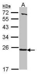
- Western Blot using HEBP1 Polyclonal Antibody (Product # PA5-30609). Sample (30 µg of whole cell lysate). Lane A: A549. 12% SDS PAGE. HEBP1 Polyclonal Antibody (Product # PA5-30609) diluted at 1:1,000.

Supportive validation
- Submitted by
- Invitrogen Antibodies (provider)
- Main image
- Experimental details
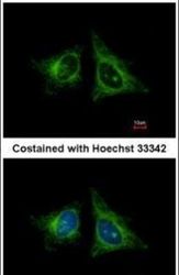
- Immunofluorescent analysis of HEBP1 in methanol-fixed HeLa cells using a HEBP1 polyclonal antibody (Product # PA5-30609) at a 1:400 dilution.

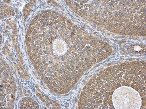
Supportive validation
- Submitted by
- Invitrogen Antibodies (provider)
- Main image
- Experimental details
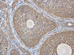
- Immunohistochemistry (Paraffin) analysis of HEBP1 was performed in paraffin-embedded mouse ovary tissue using HEBP1 Polyclonal Antibody (Product # PA5-30609) at a dilution of 1:500.
- Submitted by
- Invitrogen Antibodies (provider)
- Main image
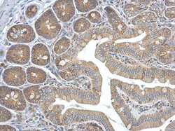
- Experimental details
- Immunohistochemistry (Paraffin) analysis of HEBP1 was performed in paraffin-embedded mouse intestine tissue using HEBP1 Polyclonal Antibody (Product # PA5-30609) at a dilution of 1:500.

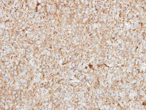
- Submitted by
- Invitrogen Antibodies (provider)
- Main image
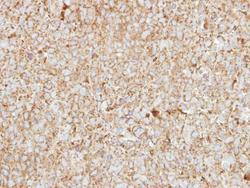
- Experimental details
- Immunohistochemical analysis of paraffin-embedded CL1-0 xenograft, using HEBP1 (Product # PA5-30609) antibody at 1:250 dilution. Antigen Retrieval: Citrate buffer, pH 6.0, 15 min.
Supportive validation
- Submitted by
- Invitrogen Antibodies (provider)
- Main image
- Experimental details
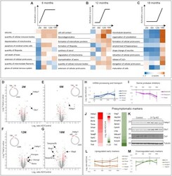
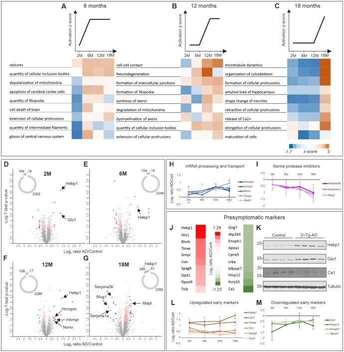
- Figure 2. Comparative proteome analysis of 3xTg-AD and control samples at different stages of AD. ( A, B, C ) Activation of biological processes at different stages of the disease assessed by Ingenuity Pathway Analysis (IPA). Heat maps represent activation z-score change over the course of disease progression and indicate pathways that are activated at 6M ( A ), 12M ( B ) and 18M ( C ). Data were obtained from four biological replicates per group for each time point. Z-score is calculated based on experimental protein expression data (log 2 AD/control ratio) and the theoretical information stored in the IPA Knowledge Base. Positive value of z-score indicates an activation of biological pathway or function. Distribution of the quantified proteins at 2M ( D ), 6M ( E ), 12M ( F ) and 18M ( G ) based on log 2 ratio AD/Control and p-value (t-test) by time point. The pie charts represent the number of quantified non-regulated proteins (grey), significantly different proteins between 3xTg-AD and control samples, t-test p-value 0.05 (pink) and significantly regulated proteins with more than 50% expression change in comparison to the control (red). ( H-I ) Dynamics of protein expression over the course of AD progression for a selection of the most regulated proteins based on their function. Proteins involved in mRNA processing and transport ( H ) that are upregulated over time and serine protease inhibitors ( I ) that are downregulated. ( J-M ) Putative presymptomatic protein markers
- Submitted by
- Invitrogen Antibodies (provider)
- Main image

- Experimental details
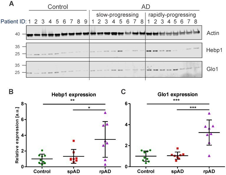
- Figure 3. Hebp1 and Glo1 exhibit increased expression in brains of patients with rapidly-progressing forms of AD. ( A ) Immunoblot analysis of Hebp1 and Glo1 expression in slow-progressing (spAD) and rapidly-progressing (rpAD) AD cases and age-matched controls. Samples from nine control, eight slow-progressing AD and eight rapidly-progressing AD patients were used in this study. Detailed information on the patients is presented in Table 2 . Quantification of ( B ) Hebp1 and ( C ) Glo1 levels in human samples. Error bars in graphs represent mean +- SD. Statistical significance in the datasets was assessed by one-way ANOVA followed by Bonferroni's multiple comparisons test for individual pairs of samples (alpha = 0.05): *p
- Submitted by
- Invitrogen Antibodies (provider)
- Main image
- Experimental details
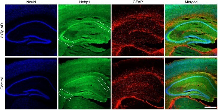
- Figure 4--figure supplement 1. Expression of Hebp1 in brains of control and 3xTg-AD mice. Immunostaining of Hebp1 in the hippocampal region of 3xTg-AD (27-month-old) and control (24-month-old) mice. Boxed regions in CA1 and dentate gyrus show representative regions where Hebp1 expression was elevated in 3xTg-AD but not control mice. Scale bar is 500 um.
- Submitted by
- Invitrogen Antibodies (provider)
- Main image
- Experimental details
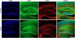
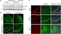
- Figure 4. Analysis of Hebp1 expression in the brain of 3xTg-AD mice. ( A ) Expression of Hebp1 in 12-month-old control and 3xTg-AD mice by brain region. ( B ) Hebp1 immunostaining of the fronto-temporal cortex depicting primary motor and somatosensory areas and hippocampus (coronal sections). CA1 region is marked with the white dashed line. ( C ) Co-staining of Hebp1 with markers of CA1 and dentate gyrus neurons (Ctip2), astrocytes (GFAP) and microglia (IBA-1) in the hippocampus of 3xTg-AD mice. Hepb1 is expressed predominantly in Ctip2-positive cells of hippocampus (neurons). All images were acquired from 12-month-old control or 3xTg-AD mice. Scale bar is 100 um. All data shown are representative of results obtained from three independent experiments. Figure 4--figure supplement 1. Expression of Hebp1 in brains of control and 3xTg-AD mice. Immunostaining of Hebp1 in the hippocampal region of 3xTg-AD (27-month-old) and control (24-month-old) mice. Boxed regions in CA1 and dentate gyrus show representative regions where Hebp1 expression was elevated in 3xTg-AD but not control mice. Scale bar is 500 um. Figure 4--figure supplement 2. Expression of Hebp1 is localized to neurons. High magnification views of the hippocampus and dentate gyrus brain regions stained with Hebp1, NeuN and GFAP. Hebp1 staining coincides with NeuN but not GFAP. Scale bar is 50 um.

- Submitted by
- Invitrogen Antibodies (provider)
- Main image
- Experimental details
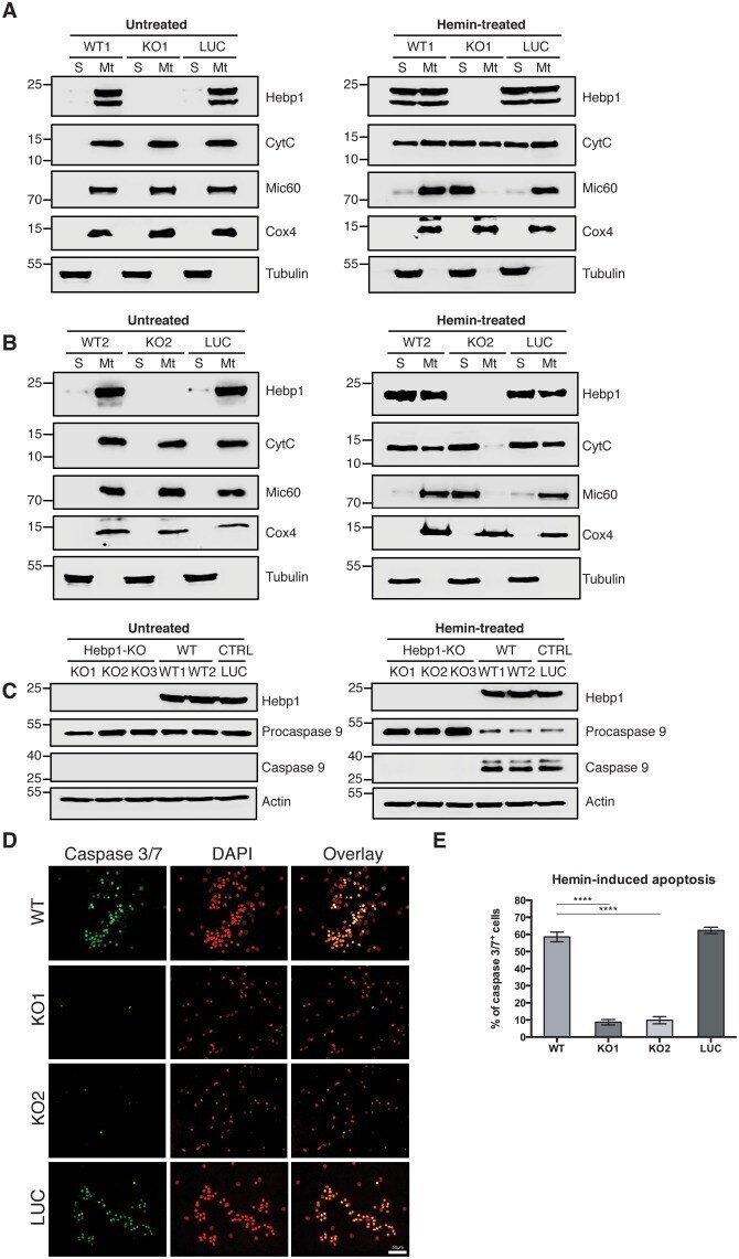
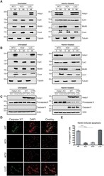
- Figure 8. Neuronal cell death occurs by triggering mitochondrial-dependent apoptotic pathway in Hebp1-expressing neurons. Western blot analyses of ( A and B ) Cyt C and Mic60 leakages and ( C ) caspase 9 activation in Hebp1-deficient, wildtype and control neurons. Wildtype and control neurons exhibited high levels of activated caspase 9 concomitant with mitochondrial release of Cyt C and Mic60 into the cytosol ( S ). Hebp1 release was also coupled with leakage of Cyt C and Mic60 in these cells. In contrast, Hebp1-deficient neurons displayed no apparent activation of caspase 9 despite leakages of Cyt C and Mic60 from neuronal mitochondria (Mt). ( D ) Wildtype, control and Hebp1-deficient neurons were treated with 10 uM hemin for 24 hr. Apoptotic cells were visualized by fluorescence staining corresponding to caspase 3/7 activation (see Materials and methods). Hebp1-deficient neurons demonstrated resistance to apoptosis upon heme overload, whereas wildtype and control neurons exhibited high levels of caspase 3/7 activity. ( E ) Quantification of the data represented by the images shown in ( D ). All bar charts represent mean +- SEM. Statistical significance in the datasets was assessed by one-way ANOVA followed by Student's t-test comparison for individual pairs of samples: ****p