Explore
 Validate
 Learn
Learn Immunocytochemistry
 Immunoprecipitation
Antibody data
- Antibody Data
- Antigen structure
- References [1]
- Comments [0]
- Validations
- Immunocytochemistry [1]
- Flow cytometry [1]
- Other assay [1]
Submit
Validation data
Reference
Comment
Report error
- Product number
- 43-8800 - Provider product page

- Provider
- Invitrogen Antibodies
- Product name
- Complex I Monoclonal Antibody (18G12BC2)
- Antibody type
- Monoclonal
- Antigen
- Other
- Description
- Positive control: fibroblasts, HL-60 cells, tissue mitochondria.
- Reactivity
- Human, Mouse, Rat, Bovine
- Host
- Mouse
- Isotype
- IgG
- Antibody clone number
- 18G12BC2
- Vial size
- 100 µg
- Concentration
- 1 mg/mL
- Storage
- 4° C, do not freeze
Submitted references TNFα drives mitochondrial stress in POMC neurons in obesity.
Yi CX, Walter M, Gao Y, Pitra S, Legutko B, Kälin S, Layritz C, García-Cáceres C, Bielohuby M, Bidlingmaier M, Woods SC, Ghanem A, Conzelmann KK, Stern JE, Jastroch M, Tschöp MH
Nature communications 2017 May 10;8:15143
Nature communications 2017 May 10;8:15143
No comments: Submit comment
Supportive validation
- Submitted by
- Invitrogen Antibodies (provider)
- Main image
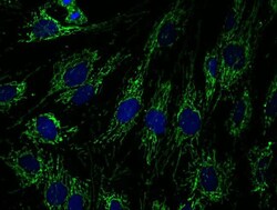
- Experimental details
- Immunocytochemistry analysis of MT-ND1 in fibroblasts cells. The cells were fixed with 4% paraformaldehyde for 20 minutes, permeabilized with 0.1% Triton X-100 for 15 minutes, and incubated with MT-ND1 Monoclonal Antibody (Product # 43-8800) at 1 µg/mL for 2 hours at room temperature. 10% Goat serum was used as the blocking agent for all blocking steps. Detection was perfomed by an Alexa Fluor® 488 goat anti-mouse IgG (H+L) at a 1:1000 dilution for 1 hour.

Supportive validation
- Submitted by
- Invitrogen Antibodies (provider)
- Main image
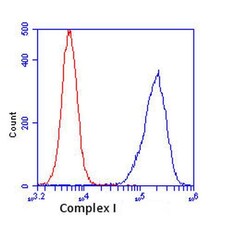
- Experimental details
- Flow cytometric analysis of MT-ND1 in HL-60 cells using a MT-ND1 Monoclonal Antibody (Product # 43-8800) at 1 µg/mL, as shown in blue. Isotype control antibody is shown in red.

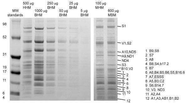
Supportive validation
- Submitted by
- Invitrogen Antibodies (provider)
- Main image
- Experimental details
- Complex I was immunopurified from mitochondria isolated from human heart (HHM), cow/bovine heart (BHM), mouse heart (MHM) and mouse brain (MBM). The lanes were stained with Coomassie Brilliant Blue R. Bands were excised from the gel and proteolytically digested for mass spectrometry analysis. For the immuno-isolation, 50 µg of Complex I Monoclonal Antibody (18G12BC2) (Product # 438800) was bound to 5 µL of swollen protein G agarose beads.