 Explore
 Validate
 Learn
Learn Western blot
Antibody data
- Antibody Data
- Antigen structure
- References [1]
- Comments [0]
- Validations
- Western blot [2]
- Immunohistochemistry [1]
- Other assay [1]
Submit
Validation data
Reference
Comment
Report error
- Product number
- PA5-77697 - Provider product page

- Provider
- Invitrogen Antibodies
- Product name
- Plexin-A1 (extracellular) Polyclonal Antibody
- Antibody type
- Polyclonal
- Antigen
- Synthetic peptide
- Description
- For reconstitution, we recommend adding 100 µL distilled water to a final antibody concentration of about 1 mg/mL. To use this carrier-free antibody for conjugation experiments, we strongly recommend performing another round of desalting. (Zeba Spin Desalting Columns, 7KMWCO, 0.5 mL, Product # 89882)
- Reactivity
- Human, Mouse, Rat
- Host
- Rabbit
- Isotype
- IgG
- Vial size
- 50 µL
- Concentration
- 0.8 mg/mL
- Storage
- -20°C
Submitted references Disruption of Sema3A/Plexin-A1 inhibitory signalling in oligodendrocytes as a therapeutic strategy to promote remyelination.
Binamé F, Pham-Van LD, Spenlé C, Jolivel V, Birmpili D, Meyer LA, Jacob L, Meyer L, Mensah-Nyagan AG, Po C, Van der Heyden M, Roussel G, Bagnard D
EMBO molecular medicine 2019 Nov 7;11(11):e10378
EMBO molecular medicine 2019 Nov 7;11(11):e10378
No comments: Submit comment
Supportive validation
- Submitted by
- Invitrogen Antibodies (provider)
- Main image

- Experimental details
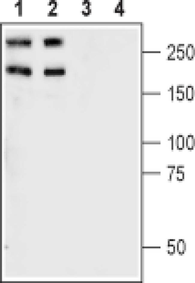
- Western blot analysis of rat (lanes 1 and 3) and mouse brain synaptosomal fraction (lanes 2 and 4) with Plexin-A1 (extracellular) polyclonal antibody (Product # PA5-77697) using a dilution of 1:400.
- Submitted by
- Invitrogen Antibodies (provider)
- Main image

- Experimental details
- Western blot analysis of rat (lanes 1 and 3) and mouse brain synaptosomal fraction (lanes 2 and 4) with Plexin-A1 (extracellular) polyclonal antibody (Product # PA5-77697) using a dilution of 1:400.

Supportive validation
- Submitted by
- Invitrogen Antibodies (provider)
- Main image

- Experimental details
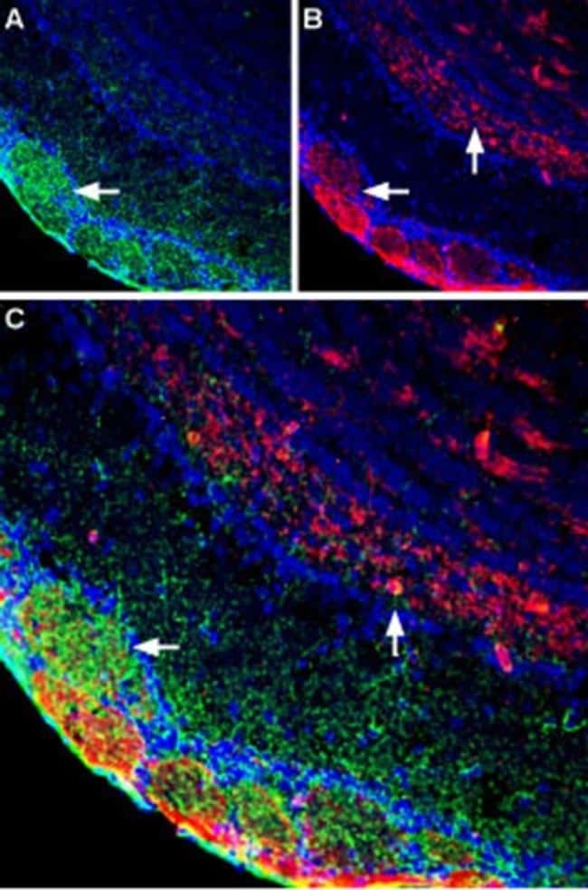
- Immunohistochemistry analysis of Plexin-A1 (extracellular) in immersion-fixed, free floating, frozen rat brain. Samples were probed with Plexin-A1 (extracellular) polyclonal antibody (Product # PA5-77697) using a dilution of 1:400, and incubated with DAPI. A) Stained glomeruli (green). B) Stained glomeruli and olfactory layers (red). Merged images also include stained nuclei.
Supportive validation
- Submitted by
- Invitrogen Antibodies (provider)
- Main image

- Experimental details
- Figure 2 Expression of Plexin-A1 in multiple sclerosis patients vs. healthy controls A-C Plexin-A1 immunoblotting analysis of brain samples of multiple sclerosis patients ( n = 11) and healthy controls ( n = 9). (A) Western blot revealed with anti-Plexin-A1 and stain-free method showing full protein content. (B) Relative expression normalized with full protein content (measured with stain-free method). Data are presented as mean +- SEM (Mann-Whitney, * P = 0.0167; n = 9 Ctrl and 11 MS patients). (C) Chi-square analysis of the proportion of patients with Plexin-A1 intensity > 2x mean control intensity. D Representative microphotographs illustrating the expression of Plexin-A1 in CNP-positive OL in healthy control or MS white matter samples (Plexin-A1: green, CNP: red). Arrowheads indicate oligodendrocytes (CNP) positive for Plexin-A1. Scale bar = 50 mum. E Quantification of the number of the CNP/Plexin-A1-positive cells in the white matter of control (HC) or MS autopsies. Data are presented as mean +- SEM (unpaired t -test, *** P = 0.0035; n = 9 Ctrl and 11 MS patients). Source data are available online for this figure.
