 Explore
 Validate
 Learn
Learn Western blot
Antibody data
- Antibody Data
- Antigen structure
- References [1]
- Comments [0]
- Validations
- Western blot [1]
- Immunocytochemistry [2]
- Immunohistochemistry [1]
- Other assay [1]
Submit
Validation data
Reference
Comment
Report error
- Product number
- PA5-38556 - Provider product page

- Provider
- Invitrogen Antibodies
- Product name
- Anti-LRAT Polyclonal Antibody
- Antibody type
- Polyclonal
- Antigen
- Synthetic peptide
- Reactivity
- Human, Mouse, Rat
- Host
- Rabbit
- Isotype
- IgG
- Vial size
- 100 µg
- Concentration
- 1 mg/mL
- Storage
- -20°C
Submitted references Vitamin A Rich Diet Diminishes Early Urothelial Carcinogenesis by Altering Retinoic Acid Signaling.
Zupančič D, Korać-Prlić J, Kreft ME, Franković L, Vilović K, Jeruc J, Romih R, Terzić J
Cancers 2020 Jun 28;12(7)
Cancers 2020 Jun 28;12(7)
No comments: Submit comment
Supportive validation
- Submitted by
- Invitrogen Antibodies (provider)
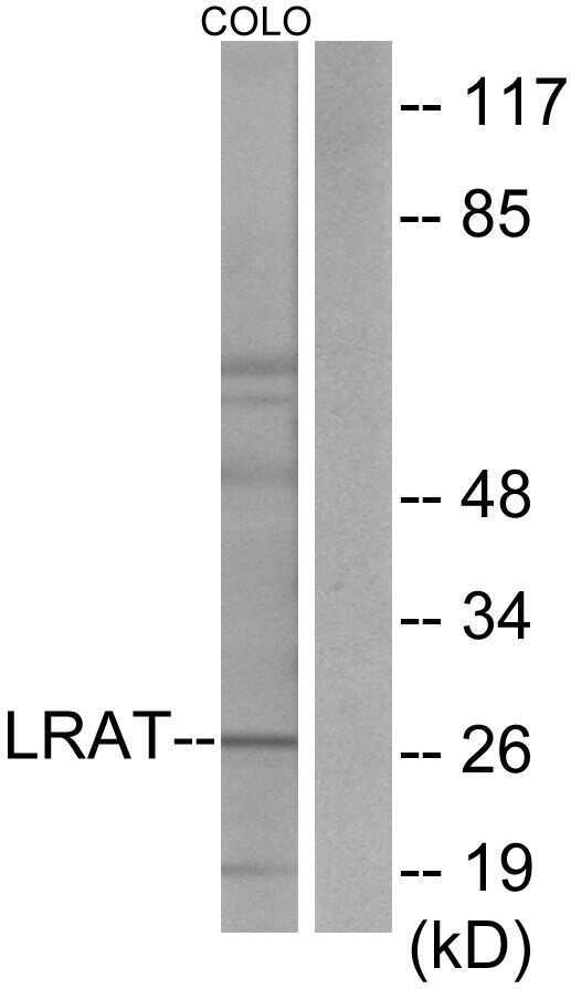
- Main image
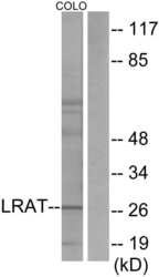
- Experimental details
- Western blot analysis of LRAT in extracts from COLO205 cells using a LRAT polyclonal antibody (Product # PA5-38556).
Supportive validation
- Submitted by
- Invitrogen Antibodies (provider)
- Main image
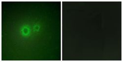
- Experimental details
- Immunofluorescent analysis of LRAT in HUVEC cells using a LRAT polyclonal antibody (Product # PA5-38556).

- Submitted by
- Invitrogen Antibodies (provider)
- Main image
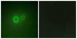
- Experimental details
- Immunofluorescent analysis of LRAT in HUVEC cells using a LRAT polyclonal antibody (Product # PA5-38556).

Supportive validation
- Submitted by
- Invitrogen Antibodies (provider)
- Main image

- Experimental details
- Immunohistochemical analysis of LRAT in paraffin-embedded human prostate carcinoma using a LRAT polyclonal antibody (Product # PA5-38556).

Supportive validation
- Submitted by
- Invitrogen Antibodies (provider)
- Main image
- Experimental details
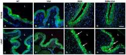
- Figure 7 Immunofluorescence of proteins stimulated by retinoic acid 6 (STRA6) and lecithin retinol acyltransferase (LRAT). ( A - D ) STRA6 labelling (green) is strong in the cytoplasm of urothelial cells of all groups. Scale bar = 50 mum. LRAT labelling (green) is present in the cytoplasm of the normal urothelial in ( E ) NT and ( F ) VitA groups. In ( G ) BBN and ( H ) BBN + VitA groups, LRAT labelling is strongest in the nuclei of urothelial cells (arrows). White line depicts the location of basal lamina, L, lumen. Scale bar = 50 mum.
