Antibody data
- Antibody Data
- Antigen structure
- References [1]
- Comments [0]
- Validations
- Western blot [2]
- Immunocytochemistry [1]
- Immunohistochemistry [1]
- Other assay [2]
Submit
Validation data
Reference
Comment
Report error
- Product number
- PA5-28510 - Provider product page

- Provider
- Invitrogen Antibodies
- Product name
- U2AF1 Polyclonal Antibody
- Antibody type
- Polyclonal
- Antigen
- Recombinant protein fragment
- Description
- Recommended positive controls: 293T, A431, H1299, HeLaS3, HepG2, Molt-4, Raji. Predicted reactivity: Mouse (99%), Rat (99%), Xenopus laevis (94%), Chicken (98%), Bovine (98%). Store product as a concentrated solution. Centrifuge briefly prior to opening the vial.
- Reactivity
- Human, Mouse
- Host
- Rabbit
- Isotype
- IgG
- Vial size
- 100 µL
- Concentration
- 1.34 mg/mL
- Storage
- Store at 4°C short term. For long term storage, store at -20°C, avoiding freeze/thaw cycles.
Submitted references FAM83F regulates canonical Wnt signalling through an interaction with CK1α.
Dunbar K, Jones RA, Dingwell K, Macartney TJ, Smith JC, Sapkota GP
Life science alliance 2021 Feb;4(2)
Life science alliance 2021 Feb;4(2)
No comments: Submit comment
Supportive validation
- Submitted by
- Invitrogen Antibodies (provider)
- Main image

- Experimental details
- Western blot analysis of U2AF35 using 30µg of A) 293T (B) A431 (C) H1299 (D) HeLa S3 (E) HepG2 (F) MOLT4 and G) Raji lysate. Samples were loaded onto a 12% SDS-PAGE gel and probed with a U2AF35 polyclonal antibody (Product # PA5-28510) at a dilution of 1:1000.
- Submitted by
- Invitrogen Antibodies (provider)
- Main image

- Experimental details
- U2AF1 Polyclonal Antibody detects U2AF35 protein by Western blot analysis. Various whole cell extracts (30 µg) were separated by 12% SDS-PAGE, and the membrane was blotted with U2AF1 Polyclonal Antibody (Product # PA5-28510) diluted by 1:1,000.
Supportive validation
- Submitted by
- Invitrogen Antibodies (provider)
- Main image

- Experimental details
- Immunofluorescent analysis of U2AF35 in paraformaldehyde-fixed HeLa cells using a U2AF35 polyclonal antibody (Product # PA5-28510) at a 1:200 dilution.
Supportive validation
- Submitted by
- Invitrogen Antibodies (provider)
- Main image

- Experimental details
- U2AF1 Polyclonal Antibody detects U2AF35 protein at nucleus on mouse intestine by immunohistochemical analysis. Sample: Paraffin-embedded mouse intestine. U2AF1 Polyclonal Antibody (Product # PA5-28510) diluted at 1:1,000. Antigen Retrieval: EDTA based buffer, pH 8.0, 15 min.
Supportive validation
- Submitted by
- Invitrogen Antibodies (provider)
- Main image

- Experimental details
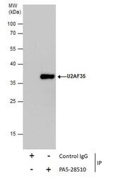
- Immunoprecipitation of U2AF35 was performed in 293T whole cell extracts using 5 µg of U2AF1 Polyclonal Antibody (Product # PA5-28510). Samples were transferred to a membrane and probed with U2AF1 Polyclonal Antibody as a primary antibody and an HRP-conjugated anti-Rabbit IgG was used as a secondary antibody.
- Submitted by
- Invitrogen Antibodies (provider)
- Main image

- Experimental details
- Figure 5. FAM83F acts upstream of glycogen synthase kinase-3beta and the loss of FAM83F protein reduces casein kinase 1alpha (CK1alpha) protein abundance at the plasma membrane. (A) qRT-PCR was performed using cDNA from HCT116 wild-type and HCT116 FAM83F -/- (cl.1) cell lines following treatment with L-CM or Wnt3A-CM with or without 0.5 muM CHIR99021 for 6 h, and primers for Axin2 and GAPDH genes. Axin2 mRNA expression was normalised to GAPDH mRNA expression and represented as arbitrary units. Data presented as scatter graph illustrating individual data points with an overlay of the mean +- SD. (B) Cytoplasmic, nuclear, and membrane lysates from HCT116 wild-type, HCT116 FAM83F -/- (cl.1), and HCT116 FAM83F -/- (cl.2) cell lines were resolved by SDS-PAGE and subjected to Western blotting with indicated antibodies. (C) Densitometry of CK1alpha protein abundance from (B) membrane lysates normalised to GAPDH protein abundance and represented as fold change compared with HCT116 wild-type cells. Data presented as scatter graph illustrating individual data points with an overlay of the mean +- SD. (D) Cytoplasmic, nuclear, and membrane lysates from DLD-1 wild-type and DLD-1 FAM83F -/- cell lines were resolved by SDS-PAGE and subjected to Western blotting with indicated antibodies. (E) Densitometry of CK1alpha protein abundance from (D) membrane lysates normalised to Na/K ATPase protein abundance and represented as fold change compared with DLD-1 wild-type cells. Data presented as sc
 Explore
 Validate
 Learn
Learn Western blot
 Immunoprecipitation