Explore
 Validate
 Learn
Learn Immunocytochemistry
Antibody data
- Antibody Data
- Antigen structure
- References [1]
- Comments [0]
- Validations
- Immunocytochemistry [1]
- Flow cytometry [1]
Submit
Validation data
Reference
Comment
Report error
- Product number
- MAB3656 - Provider product page

- Provider
- R&D Systems
- Product name
- Human Claudin-6 Antibody
- Antibody type
- Monoclonal
- Description
- Protein A or G purified from hybridoma culture supernatant. Stains human Claudin-6 transfected cells but not irrelevant transfectants.
- Reactivity
- Human
- Host
- Mouse
- Conjugate
- Unconjugated
- Antigen sequence
P56747- Isotype
- IgG
- Antibody clone number
- 342927
- Vial size
- 100 ug
- Concentration
- LYOPH
- Storage
- Use a manual defrost freezer and avoid repeated freeze-thaw cycles. 12 months from date of receipt, -20 to -70 °C as supplied. 1 month, 2 to 8 °C under sterile conditions after reconstitution. 6 months, -20 to -70 °C under sterile conditions after reconstitution.
Submitted references Identification of Novel Functions for Hepatitis C Virus Envelope Glycoprotein E1 in Virus Entry and Assembly.
Haddad JG, Rouillé Y, Hanoulle X, Descamps V, Hamze M, Dabboussi F, Baumert TF, Duverlie G, Lavie M, Dubuisson J
Journal of virology 2017 Apr 15;91(8)
Journal of virology 2017 Apr 15;91(8)
No comments: Submit comment
Supportive validation
- Submitted by
- R&D Systems (provider)
- Main image
- Experimental details
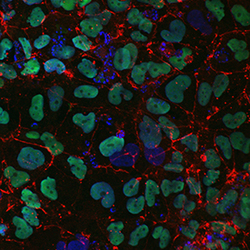
- Claudin-6 in iPS2 Human Induced Pluripotent Stem Cells. Claudin-6 was detected in immersion fixed iPS2 human induced pluripotent stem cells, differentiated into definitive endoderm with StemXVivo Endoderm Kit (Catalog # SC019), using Mouse Anti-Human Claudin-6 Monoclonal Antibody (Catalog # MAB3656) at 10 µg/mL for 3 hours at room temperature. Cells were stained using the NorthernLights™ 557-conjugated Anti-Mouse IgG Secondary Antibody (red; Catalog # NL007) and counterstained with DAPI (blue). SOX17 was also detected using Goat Anti-Human SOX17 Antigen Affinity-purified Polyclonal Antibody (green; Catalog # AF1924). Specific staining of Claudin-6 was localized to cell surfaces. View our protocol for Fluorescent ICC Staining of Stem Cells on Coverslips.

Supportive validation
- Submitted by
- R&D Systems (provider)
- Main image
- Experimental details
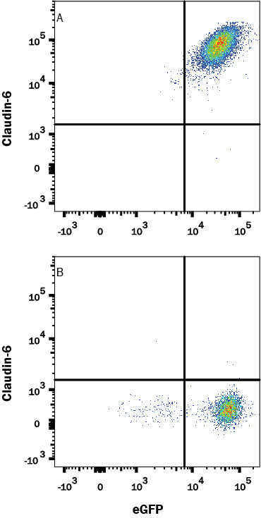
- Detection of Claudin-6 in HEK293 Human Cell Line Transfected with Human Claudin-6 and eGFP by Flow Cytometry. HEK293 human cell line transfected with (A) Human Claudin-6 or (B) irrelevant protein, and eGFP was stained with Mouse Anti-Human Claudin-6 Monoclonal Antibody (Catalog # MAB3656) followed by APC-conjugated Goat anti-Mouse IgG Secondary Antibody (Catalog # F0101B). Quadrant markers were set based on Mouse IgG2B Isotype Control Antibody (Catalog # MAB0041). View our protocol for Staining Membrane-associated Proteins.