 Explore
 Validate
 Learn
Learn Western blot
Antibody data
- Antibody Data
- Antigen structure
- References [0]
- Comments [0]
- Validations
- Western blot [2]
- Immunocytochemistry [1]
- Immunohistochemistry [1]
Submit
Validation data
Reference
Comment
Report error
- Product number
- TA328635 - Provider product page

- Provider
- OriGene
- Product name
- Rabbit Polyclonal Anti-Bombesin Receptor 2 (extracellular)
- Antibody type
- Polyclonal
- Description
- Rabbit Polyclonal Anti-Bombesin Receptor 2 (extracellular)
- Host
- Rabbit
- Conjugate
- Unconjugated
- Epitope
- GRPR
- Antibody clone number
- NULL
- Vial size
- 200 µl
- Concentration
- NULL
No comments: Submit comment
Supportive validation
- Submitted by
- OriGene (provider)
- Main image

- Experimental details
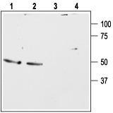
- Western blot analysis of rat (lanes 1 and 3) and mouse (lanes 2 and 4) brain lysates: 1, 2. Anti-Bombesin Receptor 2 (extracellular) antibody, (1:500). 3, 4. Anti-Bombesin Receptor 2 (extracellular) antibody, preincubated with the control peptide antigen.
- Validation comment
- WB
- Submitted by
- OriGene (provider)
- Main image

- Experimental details
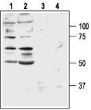
- Western blot analysis of human DU-145 (lanes 1 and 3) and PC-3 (lanes 2 and 4) prostate carcinoma cell lines: 1, 2. Anti-Bombesin Receptor 2 (extracellular) antibody, (1:200). 3, 4. Anti-Bombesin Receptor 2 (extracellular) antibody, preincubated with the control peptide antigen.
- Validation comment
- WB
Supportive validation
- Submitted by
- OriGene (provider)
- Main image

- Experimental details
- Expression of Bombesin Receptor 2 in human HT-29 cells. Immunocytochemical staining of live intact human HT-29 (colorectal adenocarcinoma) cells. Cells were stained with Anti-Bombesin Receptor 2 (extracellular) antibody(1:100), followed by goat-anti-rabbit-AlexaFluor-555 secondary antibody, showing surface expression of the BB2 receptor.
- Validation comment
- IF

Supportive validation
- Submitted by
- OriGene (provider)
- Main image
- Experimental details
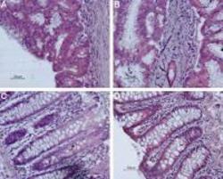
- Expression of Bombesin Receptor 2 in human colon. Immunohistochemical staining of paraffin-embedded human colon using Anti-Bombesin Receptor 2 (extracellular) antibody, (1:50). (A and B) Human colon showing malignant growth. Staining is specific for epithelium-derived malignant cells. (C and D) Normal colon, staining is specific for absorptive epithelial cells in the crypts of Lieberkuhn. Histofine (pink) is used for the color reaction. Hematoxilin is used as the counterstain.
- Validation comment
- IHC
