Explore
 Validate
 Learn
Learn Western blot
Antibody data
- Antibody Data
- Antigen structure
- References [1]
- Comments [0]
- Validations
- Western blot [3]
- Immunocytochemistry [1]
- Immunoprecipitation [1]
- Immunohistochemistry [1]
Submit
Validation data
Reference
Comment
Report error
- Product number
- GTX109736 - Provider product page

- Provider
- GeneTex
- Proper citation
- GeneTex Cat#GTX109736, RRID:AB_1949567
- Product name
- Aconitase 2 antibody [C1C3]
- Antibody type
- Polyclonal
- Reactivity
- Human, Mouse, Rat
- Host
- Rabbit
Submitted references Quercetin-induced cardioprotection against doxorubicin cytotoxicity.
Chen JY, Hu RY, Chou HC
Journal of biomedical science 2013 Dec 20;20:95
Journal of biomedical science 2013 Dec 20;20:95
No comments: Submit comment
Supportive validation
- Submitted by
- GeneTex (provider)
- Main image
- Experimental details
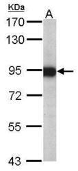
- Sample (20 ug of whole cell lysate) A: mouse brain 7.5% SDS PAGE GTX109736 diluted at 1:20000

- Submitted by
- GeneTex (provider)
- Main image
- Experimental details
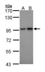
- Sample (30 ug of whole cell lysate)A: 293TB: A431 (GTX27909)7.5% SDS PAGEGTX109736 diluted at 1:10000

- Submitted by
- GeneTex (provider)
- Main image

- Experimental details
- Rat tissue extract (50 £gg) was separated by 7.5% SDS-PAGE, and the membrane was blotted with Aconitase 2 antibody [C1C3] (GTX109736) diluted at 1:10000.

Supportive validation
- Submitted by
- GeneTex (provider)
- Main image
- Experimental details
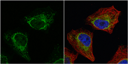
- Aconitase 2 antibody [C1C3] detects Aconitase 2 protein at mitochondria by immunofluorescent analysis.Sample: HeLa cells were fixed in 4% paraformaldehyde at RT for 15 min.Green: Aconitase 2 protein stained by Aconitase 2 antibody [C1C3] (GTX109736) diluted at 1:500.Red: alpha Tubulin, a cytoskeleton marker, stained by alpha Tubulin antibody [GT114] (GTX628802) diluted at 1:500.Blue: Hoechst 33342 staining.

Supportive validation
- Submitted by
- GeneTex (provider)
- Main image
- Experimental details
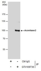
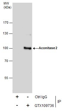
- Immunoprecipitation of Aconitase 2 protein from HeLa whole cell extracts using 5 £gg of Aconitase 2 antibody [C1C3] (GTX109736).Western blot analysis was performed using Aconitase 2 antibody [C1C3] (GTX109736).EasyBlot anti-Rabbit IgG (GTX221666-01) was used as a secondary reagent.
Supportive validation
- Submitted by
- GeneTex (provider)
- Main image
- Experimental details
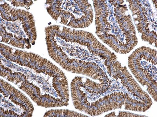
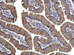
- Aconitase 2 antibody [C1C3] detects Aconitase 2 protein at mitochondria on mouse duodenum by immunohistochemical analysis. Sample: Paraffin-embedded mouse duodenum. Aconitase 2 antibody [C1C3] (GTX109736) dilution: 1:500.