Explore
 Validate
 Learn
Learn Western blot
Antibody data
- Antibody Data
- Antigen structure
- References [1]
- Comments [0]
- Validations
- Western blot [4]
- Immunocytochemistry [1]
- Immunoprecipitation [1]
- Immunohistochemistry [4]
Submit
Validation data
Reference
Comment
Report error
- Product number
- GTX114233 - Provider product page

- Provider
- GeneTex
- Proper citation
- GeneTex Cat#GTX114233, RRID:AB_10619443
- Product name
- Aconitase 2 antibody [N1N3]
- Antibody type
- Polyclonal
- Reactivity
- Human, Mouse, Rat, Chicken/Avian
- Host
- Rabbit
Submitted references The birth of quail chicks after intracytoplasmic sperm injection.
Mizushima S, Hiyama G, Shiba K, Inaba K, Dohra H, Ono T, Shimada K, Sasanami T
Development (Cambridge, England) 2014 Oct;141(19):3799-806
Development (Cambridge, England) 2014 Oct;141(19):3799-806
No comments: Submit comment
Supportive validation
- Submitted by
- GeneTex (provider)
- Main image
- Experimental details
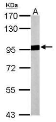
- Sample (20 ?g of whole cell lysate) A: mouse brain 7.5% SDS PAGE GTX114233 diluted at 1:10000 The HRP-conjugated anti-rabbit IgG antibody (GTX213110-01) was used to detect the primary antibody.

- Submitted by
- GeneTex (provider)
- Main image
- Experimental details
- Sample (30 ?g of whole cell lysate) A: JurKat 7.5% SDS PAGE Aconitase 2 antibody GTX114233 diluted at 1:1000 The HRP-conjugated anti-rabbit IgG antibody (GTX213110-01) was used to detect the primary antibody.

- Submitted by
- GeneTex (provider)
- Main image

- Experimental details
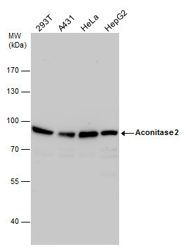
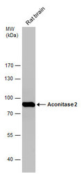
- Aconitase 2 antibody detects Aconitase 2 protein by western blot analysis. Various whole cell extracts (30 ?g) were separated by 7.5% SDS-PAGE, and the membrane was blotted with Aconitase 2 antibody (GTX114233) diluted by 1:1000. The HRP-conjugated anti-rabbit IgG antibody (GTX213110-01) was used to detect the primary antibody.
- Submitted by
- GeneTex (provider)
- Main image
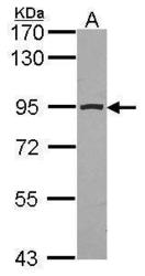
- Experimental details
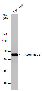
- Rat tissue extract (50 ?g) was separated by 7.5% SDS-PAGE, and the membrane was blotted with Aconitase 2 antibody [N1N3] (GTX114233) diluted at 1:10000. The HRP-conjugated anti-rabbit IgG antibody (GTX213110-01) was used to detect the primary antibody.
Supportive validation
- Submitted by
- GeneTex (provider)
- Main image

- Experimental details
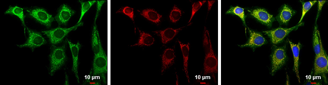
- Aconitase 2 antibody [N1N3] detects Aconitase 2 protein at mitochondria by immunofluorescent analysis.Sample: HeLa cells were fixed in 2% paraformaldehyde/culture medium at 37oC for 30 min.Green: Aconitase 2 protein stained by Aconitase 2 antibody [N1N3] (GTX114233) diluted at 1:1000.Red: MitoTrackerR Red CMXRos, a mitochondria tracker.Blue: Hoechst 33342 staining.Scale bar = 10 £gm.
Supportive validation
- Submitted by
- GeneTex (provider)
- Main image
- Experimental details
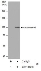
- Immunoprecipitation of Aconitase 2 protein from HeLa whole cell extracts using 5 £gg of Aconitase 2 antibody [N1N3] (GTX114233).Western blot analysis was performed using Aconitase 2 antibody [N1N3] (GTX114233).EasyBlot anti-Rabbit IgG (GTX221666-01) was used as a secondary reagent.

Supportive validation
- Submitted by
- GeneTex (provider)
- Main image
- Experimental details
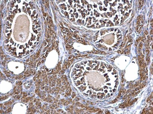
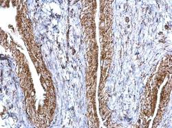
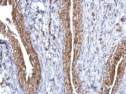
- Aconitase 2 antibody [N1N3] detects Aconitase 2 protein at mitochondria on mouse urinary bladder by immunohistochemical analysis. Sample: Paraffin-embedded mouse urinary bladder. Aconitase 2 antibody [N1N3] (GTX114233) dilution: 1:500.
- Submitted by
- GeneTex (provider)
- Main image
- Experimental details
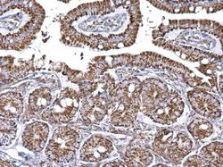
- Aconitase 2 antibody [N1N3] detects Aconitase 2 protein at mitochondria on mouse intestine by immunohistochemical analysis. Sample: Paraffin-embedded mouse intestine. Aconitase 2 antibody [N1N3] (GTX114233) dilution: 1:500.

- Submitted by
- GeneTex (provider)
- Main image
- Experimental details
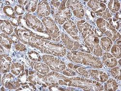
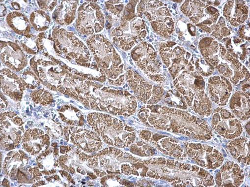
- Aconitase 2 antibody [N1N3] detects Aconitase 2 protein at mitochondria on mouse liver by immunohistochemical analysis. Sample: Paraffin-embedded mouse kidney. Aconitase 2 antibody [N1N3] (GTX114233) dilution: 1:500.
- Submitted by
- GeneTex (provider)
- Main image
- Experimental details
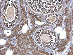
- Aconitase 2 antibody [N1N3] detects Aconitase 2 protein at mitochondria on mouse kidney by immunohistochemical analysis. Sample: Paraffin-embedded mouse ovary. Aconitase 2 antibody [N1N3] (GTX114233) dilution: 1:500.