 Explore
 Validate
 Learn
Learn Western blot
Antibody data
- Antibody Data
- Antigen structure
- References [1]
- Comments [0]
- Validations
- Western blot [2]
- Immunohistochemistry [1]
Submit
Validation data
Reference
Comment
Report error
- Product number
- AP11745PU-N - Provider product page

- Provider
- OriGene
- Product name
- Aconitase 2 (ACO2) rabbit polyclonal antibody, Purified
- Antibody type
- Polyclonal
- Description
- Aconitase 2 (ACO2) rabbit polyclonal antibody, Purified
- Host
- Rabbit
- Conjugate
- Unconjugated
- Epitope
- ACO2
- Antibody clone number
- NULL
- Vial size
- 400 µl
- Concentration
- lot specific
Submitted references Proteomic profiling reveals a severely perturbed protein expression pattern in aged skeletal muscle.
O'Connell K, Gannon J, Doran P, Ohlendieck K
International journal of molecular medicine 2007 Aug;20(2):145-53
International journal of molecular medicine 2007 Aug;20(2):145-53
No comments: Submit comment
Supportive validation
- Submitted by
- OriGene (provider)
- Main image
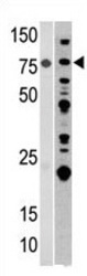
- Experimental details
- Western blot analysis of anti-ACO2 in mouse heart (left) and 293 (right) tissue lysates (35 ug/lane). ACO2 (arrow) was detected using the purified Pab (1:60 dilution).
- Validation comment
- WB

- Submitted by
- OriGene (provider)
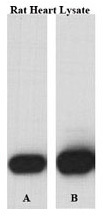
- Main image
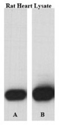
- Experimental details
- Perfused isolated rat heart whole tissue lysate was lysed with either A) 50 mM Tris-HCl, 150 mM NaCl, 1 mM EDTA, 1% NP-40, 0.1% SDS, 0.5% Na-deoxycholate, 1 mM Na3VO4, 20 mM NaF, 1 mM PMSF, 5 v/v % protease inhibitor cocktail or B) T-PER Tissue Protein Extraction Reagent, containing 1mM Na3VO4, 20 mM NaF, 5 v/v % protease inhibitor cocktail; PVDF membrane was incubated in primary Ab [rabbit polyclonal antibody against ACO2 (Center) . Solution: 1:1000 diluted in 5% NFM TBS-T 0,05 for overnight (15 hrs) at 4 ?C. Data courtesy of Boglarka Laczy M.D., Division of Cardiovascular Disease, Dept. of Medicine, University of Alabama at Birmingham.
- Validation comment
- WB
Supportive validation
- Submitted by
- OriGene (provider)
- Main image
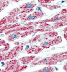
- Experimental details
- Formalin-fixed and paraffin-embedded human Heart tissue reacted with ACO2 Antibody (Center), which was peroxidase-conjugated to the secondary antibody, followed by AEC staining. This data demonstrates the use of this antibody for immunohistochemistry; clinical relevance has not been evaluated.
- Validation comment
- IHC
