 Explore
 Validate
 Learn
Learn Western blot
 Immunocytochemistry
Antibody data
- Antibody Data
- Antigen structure
- References [2]
- Comments [0]
- Validations
- Western blot [3]
- Immunoprecipitation [1]
- Immunohistochemistry [1]
Submit
Validation data
Reference
Comment
Report error
- Product number
- NBP1-32781 - Provider product page

- Provider
- Novus Biologicals
- Proper citation
- Novus Cat#NBP1-32781, RRID:AB_2221507
- Product name
- Rabbit Polyclonal Aconitase 2 Antibody
- Antibody type
- Polyclonal
- Description
- Immunogen affinity purified.
- Reactivity
- Human, Mouse, Rat
- Host
- Rabbit
- Isotype
- IgG
- Vial size
- 0.1 ml
- Storage
- Aliquot and store at -20C or -80C. Avoid freeze-thaw cycles.
Submitted references Aconitase 2 inhibits the proliferation of MCF-7 cells promoting mitochondrial oxidative metabolism and ROS/FoxO1-mediated autophagic response.
The import of the transcription factor STAT3 into mitochondria depends on GRIM-19, a component of the electron transport chain.
Ciccarone F, Di Leo L, Lazzarino G, Maulucci G, Di Giacinto F, Tavazzi B, Ciriolo MR
British journal of cancer 2020 Jan;122(2):182-193
British journal of cancer 2020 Jan;122(2):182-193
The import of the transcription factor STAT3 into mitochondria depends on GRIM-19, a component of the electron transport chain.
Tammineni P, Anugula C, Mohammed F, Anjaneyulu M, Larner AC, Sepuri NB
The Journal of biological chemistry 2013 Feb 15;288(7):4723-32
The Journal of biological chemistry 2013 Feb 15;288(7):4723-32
No comments: Submit comment
Supportive validation
- Submitted by
- Novus Biologicals (provider)
- Main image

- Experimental details
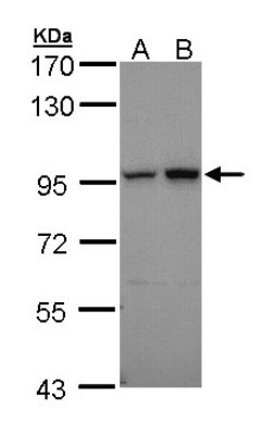
- Western Blot: Aconitase 2 Antibody [NBP1-32781] - Sample (30 ug of whole cell lysate) A: 293T B: A431 7.5% SDS PAGE, antibody diluted at 1:10000.
- Submitted by
- Novus Biologicals (provider)
- Main image

- Experimental details
- Western Blot: Aconitase 2 Antibody [NBP1-32781] - Sample (20 ug of whole cell lysate) A: mouse brain 7. 5% SDS PAGE, antibody diluted at 1:20000.

- Submitted by
- Novus Biologicals (provider)
- Main image

- Experimental details
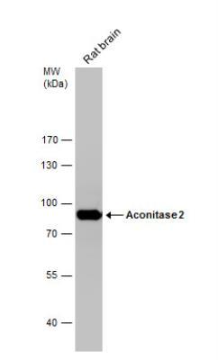
- Western Blot: Aconitase 2 Antibody [NBP1-32781] - Rat tissue extract (50 ug) was separated by 7.5% SDS-PAGE, and the membrane was blotted with Aconitase 2 antibody [C1C3] diluted at 1:10000.
Supportive validation
- Submitted by
- Novus Biologicals (provider)
- Main image

- Experimental details
- Immunoprecipitation: Aconitase 2 Antibody [NBP1-32781] - HeLa whole cell extracts using 5 ug of Aconitase 2 antibody [C1C3]. Western blot analysis was performed using Aconitase 2 antibody [C1C3]. EasyBlot anti-Rabbit IgG was used as a secondary reagent.

Supportive validation
- Submitted by
- Novus Biologicals (provider)
- Main image
- Experimental details
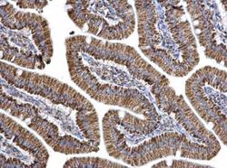
- Immunohistochemistry-Paraffin: Aconitase 2 Antibody [NBP1-32781] - Paraffin-embedded mouse duodenum. Aconitase 2 antibody [C1C3] dilution: 1:500.
