 Explore
 Validate
 Learn
Learn Western blot
 Immunocytochemistry
 Immunohistochemistry
Antibody data
- Antibody Data
- Antigen structure
- References [2]
- Comments [0]
- Validations
- Immunocytochemistry [1]
- Immunohistochemistry [4]
Submit
Validation data
Reference
Comment
Report error
- Product number
- HPA001097 - Provider product page

- Provider
- Atlas Antibodies
- Proper citation
- Atlas Antibodies Cat#HPA001097, RRID:AB_1078092
- Product name
- Anti-ACO2
- Antibody type
- Polyclonal
- Reactivity
- Human, Mouse, Rat
- Host
- Rabbit
- Conjugate
- Unconjugated
- Antigen sequence
GKKFRLEAPDADELPKGEFDPGQDTYQHPPKDSSG
QHVDVSPTSQRLQLLEPFDKWDGKDLEDLQILIKV
KGKCTTDHISAAGPWLKFRGHLDNISNNLLIGAIN
IENGKANSVRNAVTQEFGPVPDTARYYKKHGIRWV
VIGDENYGEG- Isotype
- IgG
- Vial size
- 100 µl
- Storage
- Store at +4°C for short term storage. Long time storage is recommended at -20°C.
Submitted references STED super-resolution microscopy of clinical paraffin-embedded human rectal cancer tissue.
Mitochondrial aconitase knockdown attenuates paraquat-induced dopaminergic cell death via decreased cellular metabolism and release of iron and H2O2
Ilgen P, Stoldt S, Conradi LC, Wurm CA, Rüschoff J, Ghadimi BM, Liersch T, Jakobs S
PloS one 2014;9(7):e101563
PloS one 2014;9(7):e101563
Mitochondrial aconitase knockdown attenuates paraquat-induced dopaminergic cell death via decreased cellular metabolism and release of iron and H2O2
Cantu D, Fulton R, Drechsel D, Patel M
Journal of Neurochemistry 2011 July;118(1):79-92
Journal of Neurochemistry 2011 July;118(1):79-92
No comments: Submit comment
Supportive validation
- Submitted by
- Atlas Antibodies (provider)
- Main image
- Experimental details
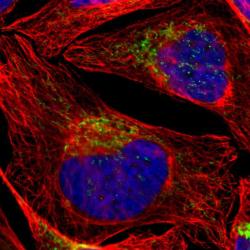
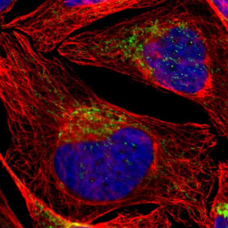
- Immunofluorescent staining of human cell line U-2 OS shows localization to mitochondria.
- Sample type
- HUMAN
Enhanced validation
Supportive validation
- Submitted by
- Atlas Antibodies (provider)
- Enhanced method
- Orthogonal validation
- Main image
- Experimental details
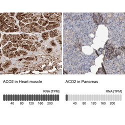
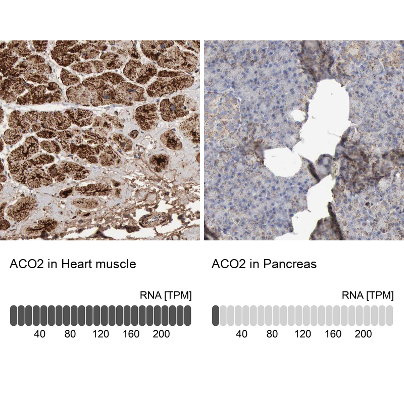
- Immunohistochemistry analysis in human heart muscle and pancreas tissues using Anti-ACO2 antibody. Corresponding ACO2 RNA-seq data are presented for the same tissues.
- Sample type
- HUMAN
Supportive validation
- Submitted by
- Atlas Antibodies (provider)
- Main image
- Experimental details
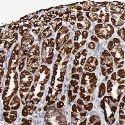
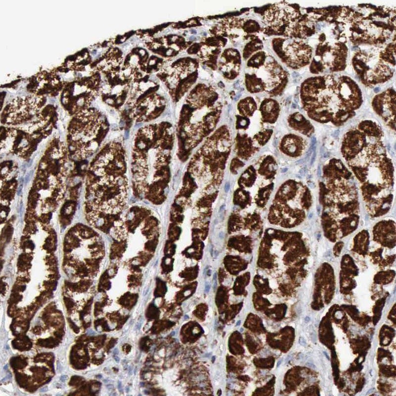
- Immunohistochemical staining of human stomach shows strong cytoplasmic positivity in glandular cells.
- Submitted by
- Atlas Antibodies (provider)
- Main image
- Experimental details
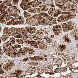
- Immunohistochemical staining of human heart muscle shows high expression.
- Sample type
- HUMAN
- Submitted by
- Atlas Antibodies (provider)
- Main image
- Experimental details
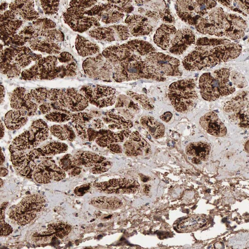
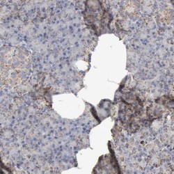
- Immunohistochemical staining of human pancreas shows low expression as expected.
- Sample type
- HUMAN
