Explore
 Validate
 Learn
Learn Western blot
 Immunoprecipitation
Antibody data
- Antibody Data
- Antigen structure
- References [0]
- Comments [0]
- Validations
- Western blot [6]
- Immunocytochemistry [2]
- Immunohistochemistry [4]
- Other assay [1]
Submit
Validation data
Reference
Comment
Report error
- Product number
- PA5-29960 - Provider product page

- Provider
- Invitrogen Antibodies
- Product name
- Aconitase 2 Polyclonal Antibody
- Antibody type
- Polyclonal
- Antigen
- Recombinant protein fragment
- Description
- Recommended positive controls: 293T, A431, HeLa, HepG2, JurKat, Mouse brain, rat brain. Predicted reactivity: Mouse (98%), Rat (97%), Zebrafish (91%), Xenopus laevis (94%), Pig (98%), Chicken (95%), Rhesus Monkey (98%), Bovine (97%). Store product as a concentrated solution. Centrifuge briefly prior to opening the vial.
- Reactivity
- Human, Mouse, Rat, Chicken/Avian
- Host
- Rabbit
- Isotype
- IgG
- Vial size
- 100 µL
- Concentration
- 1 mg/mL
- Storage
- Store at 4°C short term. For long term storage, store at -20°C, avoiding freeze/thaw cycles.
No comments: Submit comment
Supportive validation
- Submitted by
- Invitrogen Antibodies (provider)
- Main image
- Experimental details
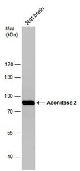
- Western Blot analysis of Aconitase 2 was performed by separating 50 µg of Rat tissue extracts by 7.5% SDS-PAGE. Proteins were transferred to a membrane and probed with a Aconitase 2 Polyclonal Antibody (Product # PA5-29960) at a dilution of 1:10000. The HRP-conjugated anti-rabbit IgG antibody was used to detect the primary antibody.
- Submitted by
- Invitrogen Antibodies (provider)
- Main image
- Experimental details
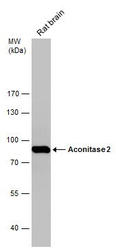
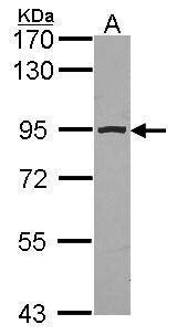
- Western Blot using Aconitase 2 Polyclonal Antibody (Product # PA5-29960). Sample (20 µg of whole cell lysate). Lane A: mouse brain. 7.5% SDS PAGE. Aconitase 2 Polyclonal Antibody (Product # PA5-29960) diluted at 1:10,000. The HRP-conjugated anti-rabbit IgG antibody was used to detect the primary antibody.

- Submitted by
- Invitrogen Antibodies (provider)
- Main image
- Experimental details
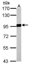
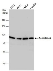
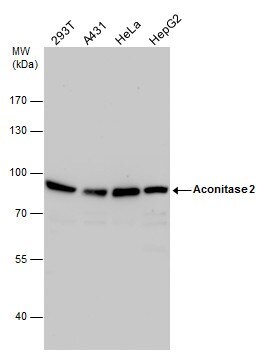
- Aconitase 2 antibody detects Aconitase 2 protein by western blot analysis. Various whole cell extracts (30 µg) were separated by 7.5% SDS-PAGE, and the membrane was blotted with Aconitase 2 antibody Aconitase 2 Polyclonal Antibody (Product # PA5-29960) diluted by 1:1,000. The HRP-conjugated anti-rabbit IgG antibody was used to detect the primary antibody.
- Submitted by
- Invitrogen Antibodies (provider)
- Main image
- Experimental details
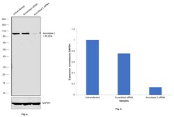
- Knockdown of Aco2; Aconitase was achieved by transfecting MCF7 cells with Aco2; Aconitase specific siRNAs (Silencer® select Product # S921, S922). Western blot analysis (Fig. a) was performed using whole cell extracts from the Aco2; Aconitase knockdown cells (lane 3), non-targeting scrambled siRNA transfected cells (lane 2) and untransfected cells (lane 1). The blot was probed with Aconitase 2 Polyclonal Antibody (Product # PA5-29960, 1:1000 dilution) and Goat anti-Rabbit IgG (H+L) Superclonal™ Recombinant Secondary Antibody, HRP (Product # A27036, 1:20,000 dilution). Densitometric analysis of this western blot is shown in histogram (Fig. b). Decrease in signal upon siRNA mediated knock down confirms that antibody is specific to Aco2; Aconitase.

- Submitted by
- Invitrogen Antibodies (provider)
- Main image

- Experimental details
- Western Blot using Aconitase 2 Polyclonal Antibody (Product # PA5-29960). Sample (30 µg of whole cell lysate). Lane A: JurKat. 7.5% SDS PAGE. Aconitase 2 antibody. Aconitase 2 Polyclonal Antibody (Product # PA5-29960) diluted at 1:1,000. The HRP-conjugated anti-rabbit IgG antibody was used to detect the primary antibody.
- Submitted by
- Invitrogen Antibodies (provider)
- Main image
- Experimental details
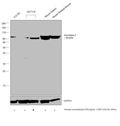
- Western blot was performed using Aconitase 2 Polyclonal Antibody (Product # PA5-29960) and a 84 kDa band corresponding to Aco2; Aconitase was observed across the cell panel tested. Induction of Aconitase was also observed upon treatment of HCT 116 cells with Human recombinant IFN alpha. Whole cell extracts (30 µg lysate) of U-2 OS (Lane 1), HCT 116 (Lane 2), HCT 116 treated with Human recombinant IFN alpha (1,000 U/mL for 24hrs) (Lane 3), Mouse Spleen (Lane 4) and Mouse Skeletal Muscle (Lane 5) were electrophoresed using NuPAGE™ 4-12% Bis-Tris Protein Gel (Product # NP0321BOX), 10 well. Resolved proteins were then transferred onto a nitrocellulose membrane (Product # IB23001) by iBlot® 2 Dry Blotting System (Product # IB21001). The blot was probed with the primary antibody (1:1000 dilution) and detected by chemiluminescence with Goat anti-Rabbit IgG (H+L) Superclonal™ Recombinant Secondary Antibody, HRP (Product # A27036, 1:20,000 dilution) using the iBright™ FL1500 Imaging System (Product # A44115). Chemiluminescent detection was performed using SuperSignal™ West Dura Extended Duration Substrate (Product # 34076).
Supportive validation
- Submitted by
- Invitrogen Antibodies (provider)
- Main image

- Experimental details
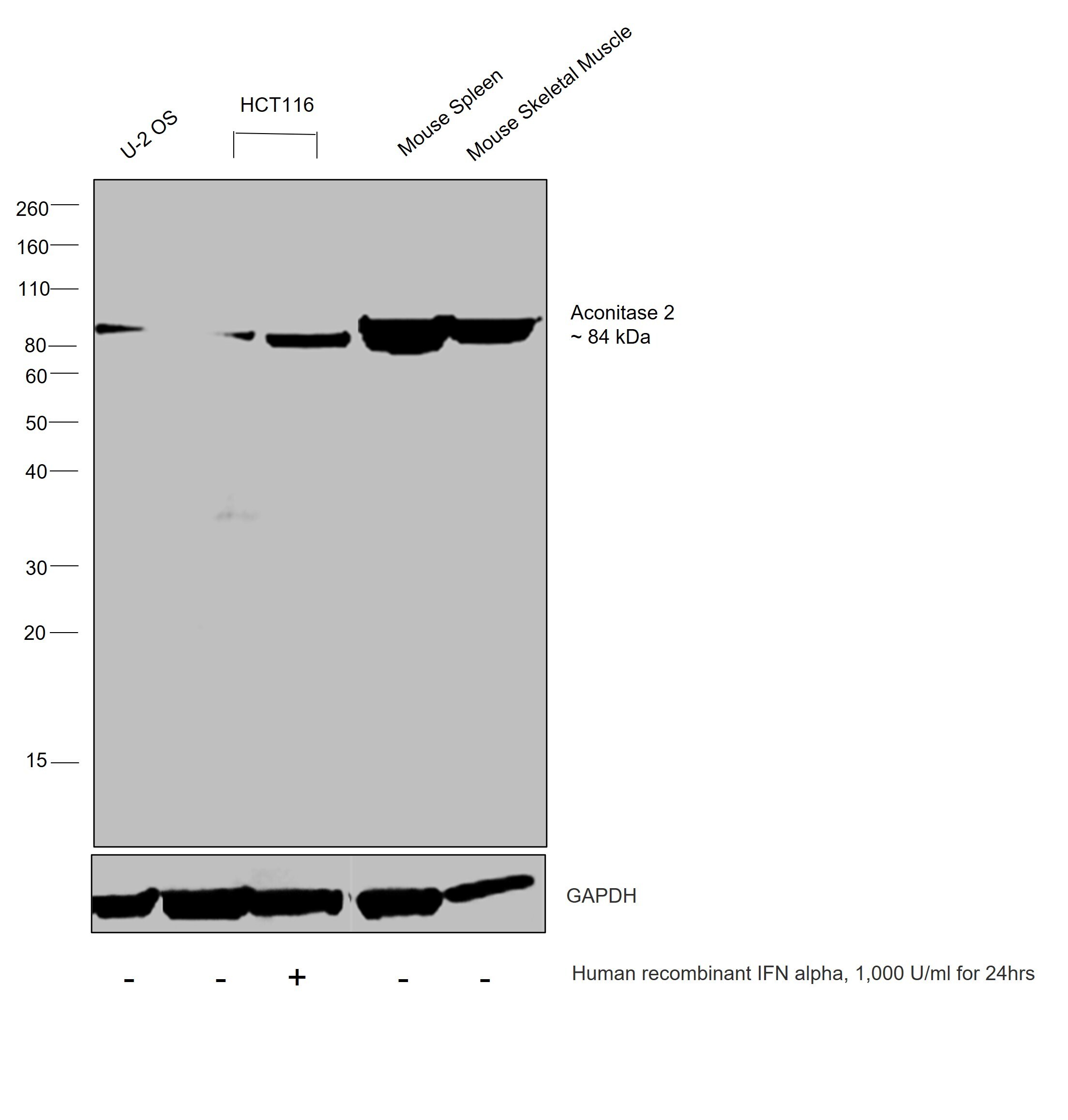
- Aconitase 2 Polyclonal Antibody detects Aconitase 2 protein at mitochondria by immunofluorescent analysis. Sample: HeLa cells were fixed in 2% paraformaldehyde/culture medium at 37oC for 30 min. Green: Aconitase 2 protein stained by Aconitase 2 Polyclonal Antibody (Product # PA5-29960) diluted at 1:1,000. Red: MitoTrackerR Red CMXRos, a mitochondria tracker. Blue: Hoechst 33342 staining. Scale bar = 10 µm.
- Submitted by
- Invitrogen Antibodies (provider)
- Main image

- Experimental details
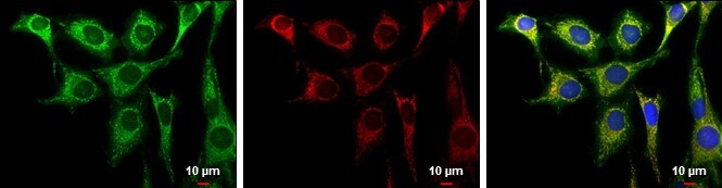
- Immunofluorescence analysis of Aco2; Aconitase was performed using 70% confluent log phase MCF7 cells. The cells were fixed with 4% paraformaldehyde for 10 minutes, permeabilized with 0.1% Triton™ X-100 for 15 minutes, and blocked with 2% BSA for 45 minutes at room temperature. The cells were labeled with Aconitase 2 Polyclonal Antibody (Product # PA5-29960) at 1:200 dilution in 0.1% BSA, incubated at 4 degree celsius overnight and then labeled with Donkey anti-Rabbit IgG (H+L) Highly Cross-Adsorbed Secondary Antibody, Alexa Fluor™ Plus 488 (Product # A32790), (1:3000 dilution), for 45 minutes at room temperature (Panel a: Green). Nuclei (Panel b:Blue) were stained with ProLong™ Diamond Antifade Mountant with DAPI (Product # P36962). F-actin (Panel c: Red) was stained with Rhodamine Phalloidin (Product # R415, 1:300). Panel d represents the merged image showing mitochondrial localization. Panel e represents control cells with no primary antibody to assess background. The images were captured at 60X magnification.
Supportive validation
- Submitted by
- Invitrogen Antibodies (provider)
- Main image
- Experimental details
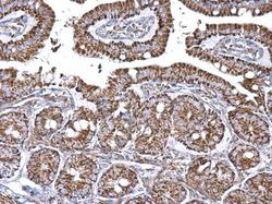
- Aconitase 2 Polyclonal Antibody detects Aconitase 2 protein at mitochondria on mouse intestine by immunohistochemical analysis. Sample: Paraffin-embedded mouse intestine. Aconitase 2 Polyclonal Antibody (Product # PA5-29960) dilution: 1:500. Antigen Retrieval: EDTA based buffer, pH 8.0, 15 min.

- Submitted by
- Invitrogen Antibodies (provider)
- Main image
- Experimental details
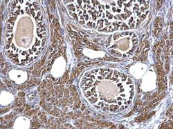
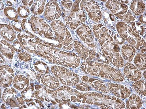
- Aconitase 2 Polyclonal Antibody detects Aconitase 2 protein at mitochondria on mouse liver by immunohistochemical analysis. Sample: Paraffin-embedded mouse kidney. Aconitase 2 Polyclonal Antibody (Product # PA5-29960) dilution: 1:500. Antigen Retrieval: EDTA based buffer, pH 8.0, 15 min.
- Submitted by
- Invitrogen Antibodies (provider)
- Main image
- Experimental details
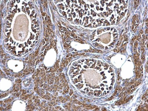
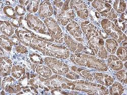
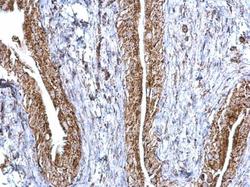
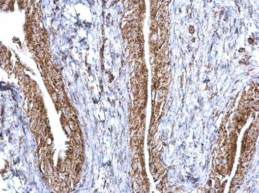
- Aconitase 2 Polyclonal Antibody detects Aconitase 2 protein at mitochondria on mouse urinary bladder by immunohistochemical analysis. Sample: Paraffin-embedded mouse urinary bladder. Aconitase 2 Polyclonal Antibody (Product # PA5-29960) dilution: 1:500. Antigen Retrieval: EDTA based buffer, pH 8.0, 15 min.
Supportive validation
- Submitted by
- Invitrogen Antibodies (provider)
- Main image
- Experimental details
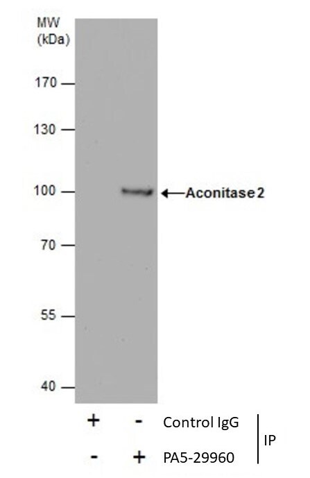
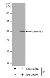
- Immunoprecipitation of Aconitase 2 was performed in HeLa whole cell extracts using 5 µg of Aconitase 2 Polyclonal Antibody (Product # PA5-29960). Samples were transferred to a membrane and probed with Aconitase 2 Polyclonal Antibody as a primary antibody and an HRP-conjugated anti-Rabbit IgG was used as a secondary antibody.