Explore
 Validate
 Learn
Learn16-5889-025
antibody from Invitrogen Antibodies
Targeting: ICOSLG
B7-H2, B7h, B7H2, B7RP-1, B7RP1, CD275, GL50, ICOS-L, ICOSL, KIAA0653
 Flow cytometry
Antibody data
- Antibody Data
- Antigen structure
- References [10]
- Comments [0]
- Validations
- Flow cytometry [1]
- Other assay [5]
Submit
Validation data
Reference
Comment
Report error
- Product number
- 16-5889-025 - Provider product page

- Provider
- Invitrogen Antibodies
- Product name
- CD275 (B7-H2) Monoclonal Antibody (MIH12), Functional Grade, eBioscience™
- Antibody type
- Monoclonal
- Antigen
- Other
- Description
- Description: The MIH12 monoclonal antibody reacts with human B7RP-1, also known as B7h, B7-H2, GL50 and ICOS Ligand. B7RP-1, a member of the B7 family, has a predicted molecular weight of approximately 40 kDa and belongs to the Ig superfamily. Human B7RP-1 is expressed by activated monocytes/macrophages. B7RP-1 binds to the ICOS molecule (AILIM, CRP-1) expressed by activated T cells. The interaction of ICOS/B7RP-1 plays an important role in the T cell costimulation pathway.
- Antibody clone number
- MIH12
- Concentration
- 1 mg/mL
Submitted references Senescence-induced endothelial phenotypes underpin immune-mediated senescence surveillance.
Cytomegalovirus restricts ICOSL expression on antigen-presenting cells disabling T cell co-stimulation and contributing to immune evasion.
Dysregulated NF-κB-Dependent ICOSL Expression in Human Dendritic Cell Vaccines Impairs T-cell Responses in Patients with Melanoma.
Comprehensive characterization of chorionic villi-derived mesenchymal stromal cells from human placenta.
Polyfunctional Melan-A-specific tumor-reactive CD8(+) T cells elicited by dacarbazine treatment before peptide-vaccination depends on AKT activation sustained by ICOS.
Development of a Novel Functional Monoclonal Antibody to Human CD275: Characterization and Biological Activity.
Pulmonary sarcoidosis is associated with high-level inducible co-stimulator (ICOS) expression on lung regulatory T cells--possible implications for the ICOS/ICOS-ligand axis in disease course and resolution.
Tumor-infiltrating plasmacytoid dendritic cells promote immunosuppression by Tr1 cells in human liver tumors.
Endothelial microparticles interact with and support the proliferation of T cells.
Expression and regulation of human CD275 on endothelial cells in healthy and inflamed mucosal tissues.
Yin K, Patten D, Gough S, de Barros Gonçalves S, Chan A, Olan I, Cassidy L, Poblocka M, Zhu H, Lun A, Schuijs M, Young A, Martinez-Jimenez C, Halim TYF, Shetty S, Narita M, Hoare M
Genes & development 2022 May 1;36(9-10):533-549
Genes & development 2022 May 1;36(9-10):533-549
Cytomegalovirus restricts ICOSL expression on antigen-presenting cells disabling T cell co-stimulation and contributing to immune evasion.
Angulo G, Zeleznjak J, Martínez-Vicente P, Puñet-Ortiz J, Hengel H, Messerle M, Oxenius A, Jonjic S, Krmpotić A, Engel P, Angulo A
eLife 2021 Jan 18;10
eLife 2021 Jan 18;10
Dysregulated NF-κB-Dependent ICOSL Expression in Human Dendritic Cell Vaccines Impairs T-cell Responses in Patients with Melanoma.
Maurer DM, Adamik J, Santos PM, Shi J, Shurin MR, Kirkwood JM, Storkus WJ, Butterfield LH
Cancer immunology research 2020 Dec;8(12):1554-1567
Cancer immunology research 2020 Dec;8(12):1554-1567
Comprehensive characterization of chorionic villi-derived mesenchymal stromal cells from human placenta.
Ventura Ferreira MS, Bienert M, Müller K, Rath B, Goecke T, Opländer C, Braunschweig T, Mela P, Brümmendorf TH, Beier F, Neuss S
Stem cell research & therapy 2018 Feb 5;9(1):28
Stem cell research & therapy 2018 Feb 5;9(1):28
Polyfunctional Melan-A-specific tumor-reactive CD8(+) T cells elicited by dacarbazine treatment before peptide-vaccination depends on AKT activation sustained by ICOS.
Franzese O, Palermo B, Di Donna C, Sperduti I, Ferraresi V, Stabile H, Gismondi A, Santoni A, Nisticò P
Oncoimmunology 2016 May;5(5):e1114203
Oncoimmunology 2016 May;5(5):e1114203
Development of a Novel Functional Monoclonal Antibody to Human CD275: Characterization and Biological Activity.
Hu X, Liu C, An J, Shen Y, Hu Y, Jiang J, Wu J, Zhang X
Monoclonal antibodies in immunodiagnosis and immunotherapy 2016 Feb;35(1):18-24
Monoclonal antibodies in immunodiagnosis and immunotherapy 2016 Feb;35(1):18-24
Pulmonary sarcoidosis is associated with high-level inducible co-stimulator (ICOS) expression on lung regulatory T cells--possible implications for the ICOS/ICOS-ligand axis in disease course and resolution.
Sakthivel P, Grunewald J, Eklund A, Bruder D, Wahlström J
Clinical and experimental immunology 2016 Feb;183(2):294-306
Clinical and experimental immunology 2016 Feb;183(2):294-306
Tumor-infiltrating plasmacytoid dendritic cells promote immunosuppression by Tr1 cells in human liver tumors.
Pedroza-Gonzalez A, Zhou G, Vargas-Mendez E, Boor PP, Mancham S, Verhoef C, Polak WG, Grünhagen D, Pan Q, Janssen H, Garcia-Romo GS, Biermann K, Tjwa ET, IJzermans JN, Kwekkeboom J, Sprengers D
Oncoimmunology 2015 Jun;4(6):e1008355
Oncoimmunology 2015 Jun;4(6):e1008355
Endothelial microparticles interact with and support the proliferation of T cells.
Wheway J, Latham SL, Combes V, Grau GE
Journal of immunology (Baltimore, Md. : 1950) 2014 Oct 1;193(7):3378-87
Journal of immunology (Baltimore, Md. : 1950) 2014 Oct 1;193(7):3378-87
Expression and regulation of human CD275 on endothelial cells in healthy and inflamed mucosal tissues.
Youngnak-Piboonratanakit P, Tsushima F, Otsuki N, Igarashi H, Omura K, Azuma M
Scandinavian journal of immunology 2006 Mar;63(3):191-8
Scandinavian journal of immunology 2006 Mar;63(3):191-8
No comments: Submit comment
Supportive validation
- Submitted by
- Invitrogen Antibodies (provider)
- Main image
- Experimental details
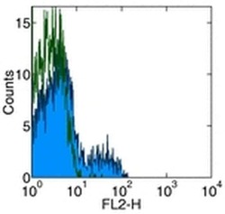
- Staining of normal human peripheral blood cells with Anti-Human CD275 (B7-H2) PE.Appropriate isotype controls were used (open histogram).Cells in the lymphocyte population were used for analysis.

Supportive validation
- Submitted by
- Invitrogen Antibodies (provider)
- Main image
- Experimental details
- NULL

- Submitted by
- Invitrogen Antibodies (provider)
- Main image
- Experimental details
- NULL

- Submitted by
- Invitrogen Antibodies (provider)
- Main image

- Experimental details
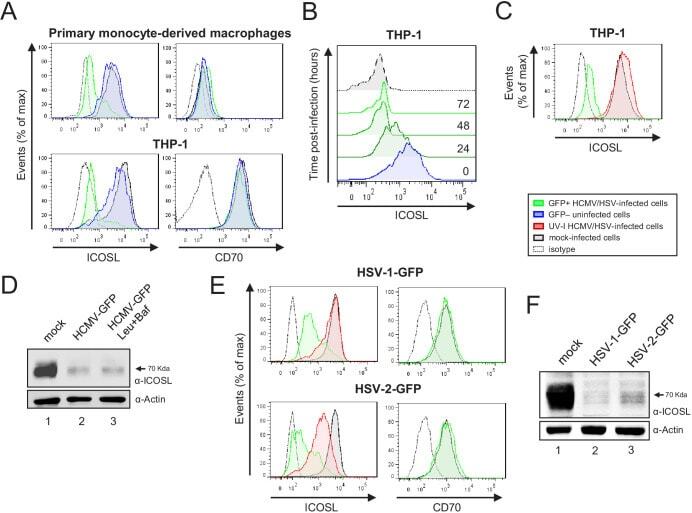
- Figure 11. HCMV, HSV-1, and HSV-2 also limit cell surface expression of ICOSL on antigen-presenting cells (APCs). ( A ) Primary monocyte-derived macrophages (upper panels) and PMA-treated THP-1 cells (bottom panels) were mock-infected or infected for 72 hr with HCMV-GFP at an moi of 10 and analyzed by flow cytometry for cell-surface expression of human ICOSL or CD70 using specific mAbs against each of these receptors. Gray histograms represent the expression of mock-infected cells, green histograms represent the expression on HCMV-infected (GFP+) cells, and blue histograms represent the expression on uninfected (GFP-) cells from the same culture. ( B ) PMA-treated THP-1 cells were mock-infected (time 0) or infected with HCMV-GFP as in A and analyzed by flow cytometry for surface expression of ICOSL at the different time points after infection indicated. ( C ) Same as in A, except that an moi of 20 was used, and THP-1 cells were also exposed for 72 hr to the same amount of HCMV-GFP UV-inactivated (red histogram). ( D ) Equal amounts of lysates from PMA-treated THP-1 cells mock-infected (lane 1) or infected for 72 hr at an moi of 20 with HCMV-GFP (lanes 2 and 3), and when indicated, treated with 250 muM leupeptin and 20 nM of bafilomycin A1 (lane 3), were lysed and analyzed by western blot with antibodies against ICOSL and actin, followed by anti-rabbit IgG-HRP (ICOSL) or anti-mouse IgG-HRP (actin). ( E ) PMA-treated THP-1 cells were mock-infected or infected with HSV-1-GFP, HS
- Submitted by
- Invitrogen Antibodies (provider)
- Main image
- Experimental details
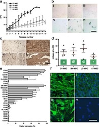
- Fig. 2 a Cumulative population-doubling (cPD) levels versus passage number for the four different sources of MSC. Black represents CV-MSC ( n = 7), dark gray UC-MSC ( n = 4), medium gray AT-MSC ( n = 5), and light gray BM-MSC ( n = 6). b IHC-based senescence-associated beta-galactosidase (SA-beta-gal) staining of CV-MSC in early (i, passage 4) and late (ii, passage 9) passages, AT-MSC in passage 6 (iii), BM-MSC in passage 6 (iv), and UC-MSC in passage 2 (v) and passage 4 (vi). Scale = 200 mum. c IHC of CV-MSC (i, ii) and BM-MSC (iii, iv) stained for osteopontin (i, iii) and fibronectin (ii, iv). Scale = 1 mm. d Collagen area (%) after collagen contraction assay for CV-MSC ( n = 4), BM-MSC ( n = 3), UC-MSC ( n = 4), and AT-MSC ( n = 3). Cells in passage 3 were used. Results expressed as mean +- SD, percentage of the total collagen area of the collagen gels without cells. e Surface marker expression of CV-MSC in early passages ( n = 5). Results expressed as mean +- SD (%). f Representative immunofluorescence of early passaged CV-MSC (i, iii) and BM-MSC (iii, iv) stained for SM22alpha (i, iii) and alpha-SMA (ii, iv). Scale = 50 mum. AT adipose tissue, BM bone marrow, CV chorionic villi, MSC mesenchymal stromal cells, UC umbilical cord
- Submitted by
- Invitrogen Antibodies (provider)
- Main image
- Experimental details
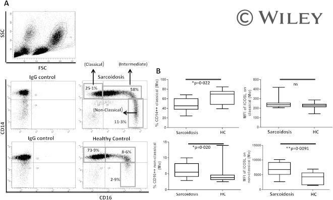
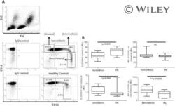
- Increased proportion of non-classical blood monocytes with higher inducible co-stimulator ligand (ICOS-L) expression in sarcoidosis patients. (a) Representative fluorescence activated cell sorter (FACS) dot-plot gated on blood monocyte populations in peripheral blood mononuclear cells (PBMCs) showing the frequencies of classical, intermediate and non-classical (proinflammatory) monocytes in sarcoidosis patients ( n = 11) and healthy controls ( n = 13). (b) Box-plots represent the % CD14 high CD16 low monocytes (classical) in sarcoidosis patients and healthy controls (upper left) and the intensities of ICOS-L [as mean fluorescence intensity (MFI)] on classical monocytes in patients and controls (upper right). Box-plots represent the % CD14 low CD16 high monocytes (non-classical) in sarcoidosis patients and healthy controls (lower left) and the intensities of ICOS-L (as MFI) on non-classical monocytes in patients and controls (lower right). P -values were calculated using the Mann-Whitney U -test.