Antibody data
- Antibody Data
- Antigen structure
- References [12]
- Comments [0]
- Validations
- Western blot [9]
- Immunocytochemistry [3]
- Immunoprecipitation [1]
- Immunohistochemistry [3]
Submit
Validation data
Reference
Comment
Report error
- Product number
- GTX109669 - Provider product page

- Provider
- GeneTex
- Proper citation
- GeneTex Cat#GTX109669, RRID:AB_1949824
- Product name
- Calnexin antibody [C3], C-term
- Antibody type
- Polyclonal
- Reactivity
- Human, Mouse, Rat, Sheep
- Host
- Rabbit
Submitted references Pigment epithelium-derived factor inhibits lung cancer migration and invasion by upregulating exosomal thrombospondin 1.
A high-throughput pipeline for validation of antibodies.
Selective serotonin reuptake inhibitor, fluoxetine, impairs E-cadherin-mediated cell adhesion and alters calcium homeostasis in pancreatic beta cells.
Phosphoinositide phosphatase Sac3 regulates the cell surface expression of scavenger receptor A and formation of lipid droplets in macrophages.
A familial ATP13A2 mutation enhances alpha-synuclein aggregation and promotes cell death.
Suppression of Cytochrome P450 3A4 Function by UDP-Glucuronosyltransferase 2B7 through a Protein-Protein Interaction: Cooperative Roles of the Cytosolic Carboxyl-Terminal Domain and the Luminal Anchoring Region.
Recent studies of ovine neuronal ceroid lipofuscinoses from BARN, the Batten Animal Research Network.
APOBEC3A cytidine deaminase induces RNA editing in monocytes and macrophages.
Deficient nitric oxide signalling impairs skeletal muscle growth and performance: involvement of mitochondrial dysregulation.
Cadmium-based quantum dot induced autophagy formation for cell survival via oxidative stress.
R26R-GR: a Cre-activable dual fluorescent protein reporter mouse.
Saturated fatty acids induce c-Src clustering within membrane subdomains, leading to JNK activation.
Huang WT, Chong IW, Chen HL, Li CY, Hsieh CC, Kuo HF, Chang CY, Chen YH, Liu YP, Lu CY, Liu YR, Liu PL
Cancer letters 2019 Feb 1;442:287-298
Cancer letters 2019 Feb 1;442:287-298
A high-throughput pipeline for validation of antibodies.
Sikorski K, Mehta A, Inngjerdingen M, Thakor F, Kling S, Kalina T, Nyman TA, Stensland ME, Zhou W, de Souza GA, Holden L, Stuchly J, Templin M, Lund-Johansen F
Nature methods 2018 Nov;15(11):909-912
Nature methods 2018 Nov;15(11):909-912
Selective serotonin reuptake inhibitor, fluoxetine, impairs E-cadherin-mediated cell adhesion and alters calcium homeostasis in pancreatic beta cells.
Chang HY, Chen SL, Shen MR, Kung ML, Chuang LM, Chen YW
Scientific reports 2017 Jun 14;7(1):3515
Scientific reports 2017 Jun 14;7(1):3515
Phosphoinositide phosphatase Sac3 regulates the cell surface expression of scavenger receptor A and formation of lipid droplets in macrophages.
Morioka S, Nigorikawa K, Hazeki K, Ohmura M, Sakamoto H, Matsumura T, Takasuga S, Hazeki O
Experimental cell research 2017 Aug 15;357(2):252-259
Experimental cell research 2017 Aug 15;357(2):252-259
A familial ATP13A2 mutation enhances alpha-synuclein aggregation and promotes cell death.
Lopes da Fonseca T, Pinho R, Outeiro TF
Human molecular genetics 2016 Jul 15;25(14):2959-2971
Human molecular genetics 2016 Jul 15;25(14):2959-2971
Suppression of Cytochrome P450 3A4 Function by UDP-Glucuronosyltransferase 2B7 through a Protein-Protein Interaction: Cooperative Roles of the Cytosolic Carboxyl-Terminal Domain and the Luminal Anchoring Region.
Miyauchi Y, Nagata K, Yamazoe Y, Mackenzie PI, Yamada H, Ishii Y
Molecular pharmacology 2015 Oct;88(4):800-12
Molecular pharmacology 2015 Oct;88(4):800-12
Recent studies of ovine neuronal ceroid lipofuscinoses from BARN, the Batten Animal Research Network.
Palmer DN, Neverman NJ, Chen JZ, Chang CT, Houweling PJ, Barry LA, Tammen I, Hughes SM, Mitchell NL
Biochimica et biophysica acta 2015 Oct;1852(10 Pt B):2279-86
Biochimica et biophysica acta 2015 Oct;1852(10 Pt B):2279-86
APOBEC3A cytidine deaminase induces RNA editing in monocytes and macrophages.
Sharma S, Patnaik SK, Taggart RT, Kannisto ED, Enriquez SM, Gollnick P, Baysal BE
Nature communications 2015 Apr 21;6:6881
Nature communications 2015 Apr 21;6:6881
Deficient nitric oxide signalling impairs skeletal muscle growth and performance: involvement of mitochondrial dysregulation.
De Palma C, Morisi F, Pambianco S, Assi E, Touvier T, Russo S, Perrotta C, Romanello V, Carnio S, Cappello V, Pellegrino P, Moscheni C, Bassi MT, Sandri M, Cervia D, Clementi E
Skeletal muscle 2014;4(1):22
Skeletal muscle 2014;4(1):22
Cadmium-based quantum dot induced autophagy formation for cell survival via oxidative stress.
Luo YH, Wu SB, Wei YH, Chen YC, Tsai MH, Ho CC, Lin SY, Yang CS, Lin P
Chemical research in toxicology 2013 May 20;26(5):662-73
Chemical research in toxicology 2013 May 20;26(5):662-73
R26R-GR: a Cre-activable dual fluorescent protein reporter mouse.
Chen YT, Tsai MS, Yang TL, Ku AT, Huang KH, Huang CY, Chou FJ, Fan HH, Hong JB, Yen ST, Wang WL, Lin CC, Hsu YC, Su KY, Su IC, Jang CW, Behringer RR, Favaro R, Nicolis SK, Chien CL, Lin SW, Yu IS
PloS one 2012;7(9):e46171
PloS one 2012;7(9):e46171
Saturated fatty acids induce c-Src clustering within membrane subdomains, leading to JNK activation.
Holzer RG, Park EJ, Li N, Tran H, Chen M, Choi C, Solinas G, Karin M
Cell 2011 Sep 30;147(1):173-84
Cell 2011 Sep 30;147(1):173-84
No comments: Submit comment
Enhanced validation
Supportive validation
- Submitted by
- GeneTex (provider)
- Enhanced method
- Genetic validation
- Main image
- Experimental details
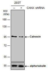
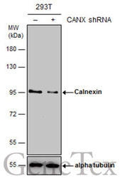
- Non-transfected (¡V) and transfected (+) 293T whole cell extracts (15 ?g) were separated by 7.5% SDS-PAGE, and the membrane was blotted with Calnexin antibody [C3], C-term (GTX109669) diluted at 1:20000. The HRP-conjugated anti-rabbit IgG antibody (GTX213110-01) was used to detect the primary antibody.
Supportive validation
- Submitted by
- GeneTex (provider)
- Main image

- Experimental details
- Sample (30 ug of whole cell lysate) A: 293T B: A431 C: HeLa D: HepG2 7.5% SDS PAGE GTX109669 diluted at 1:10000
- Validation comment
- WB
- Submitted by
- GeneTex (provider)
- Main image
- Experimental details
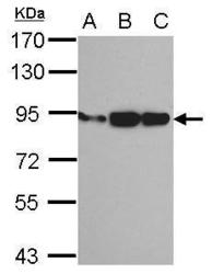
- Sample (30 ?g of whole cell lysate) A: NIH-3T3 B: JC C: BCL-1 7.5% SDS PAGE GTX109669 diluted at 1:1000 The HRP-conjugated anti-rabbit IgG antibody (GTX213110-01) was used to detect the primary antibody.
- Submitted by
- GeneTex (provider)
- Main image
- Experimental details
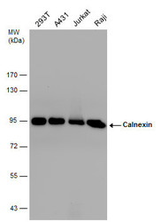
- Calnexin antibody detects Calnexin protein by western blot analysis. Various whole cell extracts (30 ?g) were separated by 7.5% SDS-PAGE, and the membrane was blotted with Calnexin antibody (GTX109669) diluted by 1:10000. The HRP-conjugated anti-rabbit IgG antibody (GTX213110-01) was used to detect the primary antibody.
- Submitted by
- GeneTex (provider)
- Main image
- Experimental details
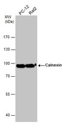
- Various whole cell extracts (30 ?g) were separated by 7.5% SDS-PAGE, and the membrane was blotted with Calnexin antibody (GTX109669) diluted at 1:1000. The HRP-conjugated anti-rabbit IgG antibody (GTX213110-01) was used to detect the primary antibody.
- Submitted by
- GeneTex (provider)
- Main image
- Experimental details
- Non-transfected (¡V) and transfected (+) 293T whole cell extracts (15 ?g) were separated by 7.5% SDS-PAGE, and the membrane was blotted with Calnexin antibody [C3], C-term (GTX109669) diluted at 1:20000. The HRP-conjugated anti-rabbit IgG antibody (GTX213110-01) was used to detect the primary antibody.
- Submitted by
- GeneTex (provider)
- Main image
- Experimental details
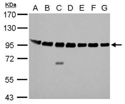
- Calnexin antibody [C3], C-term detects Calnexin protein by Western blot analysis.A. 30 £gg Neuro2A whole cell lysate/extractB. 30 £gg GL261 whole cell lysate/extractC. 30 £gg C8D30 whole cell lysate/extractD. 30 £gg NIH-3T3 whole cell lysate/extractE. 30 £gg BCL-1 whole cell lysate/extractF. 30 £gg Raw264.7 whole cell lysate/extractG. 30 £gg C2C12 whole cell lysate/extract7.5 % SDS-PAGECalnexin antibody [C3], C-term (GTX109669) dilution: 1:10000
- Submitted by
- GeneTex (provider)
- Main image
- Experimental details
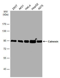
- Various whole cell extracts (30 ?g) were separated by 7.5% SDS-PAGE, and the membrane was blotted with Calnexin antibody [C3], C-term (GTX109669) diluted at 1:10000. The HRP-conjugated anti-rabbit IgG antibody (GTX213110-01) was used to detect the primary antibody.
- Submitted by
- GeneTex (provider)
- Main image
- Experimental details
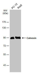
- Various whole cell extracts (30 ?g) were separated by 7.5% SDS-PAGE, and the membrane was blotted with Calnexin antibody [C3], C-term (GTX109669) diluted at 1:10000. The HRP-conjugated anti-rabbit IgG antibody (GTX213110-01) was used to detect the primary antibody.
Supportive validation
- Submitted by
- GeneTex (provider)
- Main image

- Experimental details
- Confocal immunofluorescence analysis (Olympus FV10i) of methanol-fixed HeLa, using Calnexin(GTX109669) antibody (Green) at 1:500 dilution. Alpha-tubulin filaments were labeled with GTX11304 (Red) at 1:2500.
- Submitted by
- GeneTex (provider)
- Main image
- Experimental details
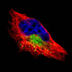
- Confocal immunofluorescence analysis (Olympus FV10i) of paraformaldehyde-fixed HeLa, using Calnexin(GTX109669) antibody (Green) at 1:200 dilution. Alpha-tubulin filaments were labeled with GTX11304 (Red) at 1:2500.
- Submitted by
- GeneTex (provider)
- Main image
- Experimental details
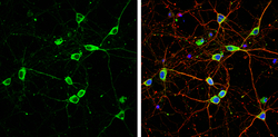
- Calnexin antibody [C3], C-term detects Calnexin protein by immunofluorescent analysis.Sample: DIV9 rat E18 primary cortical neuron cells were fixed in 4% paraformaldehyde at RT for 15 min.Green: Calnexin stained by Calnexin antibody [C3], C-term (GTX109669) diluted at 1:500.Red: beta Tubulin 3/ Tuj1, stained by beta Tubulin 3/ Tuj1 antibody [GT1338] (GTX631831) diluted at 1:500.Blue: Fluoroshield with DAPI (GTX30920).
Supportive validation
- Submitted by
- GeneTex (provider)
- Main image

- Experimental details
- Calnexin antibody immunoprecipitates Calnexin protein in IP experiments. IP Sample: 1000 ?g HeLa whole cell lysate/extract A. 30 £gg HeLa whole cell lysate/extract B. Control with 2 £gg of preimmune rabbit IgG C. Immunoprecipitation of Calnexin protein by 2 £gg of Calnexin antibody (GTX109669) 7.5% SDS-PAGE The immunoprecipitated Calnexin protein was detected by Calnexin antibody (GTX109669) diluted at 1:1000. EasyBlot anti-rabbit IgG (GTX221666-01) was used as a secondary reagent.
Supportive validation
- Submitted by
- GeneTex (provider)
- Main image
- Experimental details
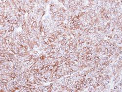
- Immunohistochemical analysis of paraffin-embedded DLD-1 xenograft, using Calnexin(GTX109669) antibody at 1:500 dilution.
- Submitted by
- GeneTex (provider)
- Main image
- Experimental details
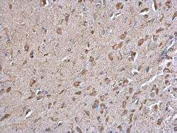
- Calnexin antibody [C3], C-term detects Calnexin protein at cytosol on mouse middle brain by immunohistochemical analysis. Sample: Paraffin-embedded mouse middle brain. Calnexin antibody [C3], C-term (GTX109669) dilution: 1:500.
- Submitted by
- GeneTex (provider)
- Main image
- Experimental details
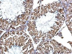
- Calnexin antibody [C3], C-term detects Calnexin protein at cytosol on mouse testis by immunohistochemical analysis. Sample: Paraffin-embedded mouse testis. Calnexin antibody [C3], C-term (GTX109669) dilution: 1:500.
 Explore
 Validate
 Learn
Learn Western blot
 Immunocytochemistry