Explore
 Validate
 Learn
Learn Western blot
Antibody data
- Antibody Data
- Antigen structure
- References [5]
- Comments [0]
- Validations
- Western blot [7]
- Immunocytochemistry [1]
- Immunohistochemistry [7]
- Other assay [4]
Submit
Validation data
Reference
Comment
Report error
- Product number
- PA5-34665 - Provider product page

- Provider
- Invitrogen Antibodies
- Product name
- Calnexin Polyclonal Antibody
- Antibody type
- Polyclonal
- Antigen
- Recombinant protein fragment
- Description
- Recommended positive controls: 293T, A431, H1299, HeLaS3, HepG2, Molt-4, Raji, NIH-3T3, PC-12, Rat2.
- Concentration
- 0.16 mg/mL
Submitted references Protamine Sulfate Is a Potent Inhibitor of Human Papillomavirus Infection In Vitro and In Vivo.
Deciphering the Role of Extracellular Vesicles Derived from ZIKV-Infected hcMEC/D3 Cells on the Blood-Brain Barrier System.
Regulation of intestinal epithelial intercellular adhesion and barrier function by desmosomal cadherin desmocollin-2.
The D614G Mutation Enhances the Lysosomal Trafficking of SARS-CoV-2 Spike.
Optimization of Morpholino Antisense Oligonucleotides Targeting the Intronic Repressor Element1 in Spinal Muscular Atrophy.
Young JM, Zine El Abidine A, Gómez-Martinez RA, Bondu V, Sterk RT, Surviladze Z, Ozbun MA
Antimicrobial agents and chemotherapy 2022 Jan 18;66(1):e0151321
Antimicrobial agents and chemotherapy 2022 Jan 18;66(1):e0151321
Deciphering the Role of Extracellular Vesicles Derived from ZIKV-Infected hcMEC/D3 Cells on the Blood-Brain Barrier System.
Fikatas A, Dehairs J, Noppen S, Doijen J, Vanderhoydonc F, Meyen E, Swinnen JV, Pannecouque C, Schols D
Viruses 2021 Nov 25;13(12)
Viruses 2021 Nov 25;13(12)
Regulation of intestinal epithelial intercellular adhesion and barrier function by desmosomal cadherin desmocollin-2.
Raya-Sandino A, Luissint AC, Kusters DHM, Narayanan V, Flemming S, Garcia-Hernandez V, Godsel LM, Green KJ, Hagen SJ, Conway DE, Parkos CA, Nusrat A
Molecular biology of the cell 2021 Apr 15;32(8):753-768
Molecular biology of the cell 2021 Apr 15;32(8):753-768
The D614G Mutation Enhances the Lysosomal Trafficking of SARS-CoV-2 Spike.
Guo C, Tsai SJ, Ai Y, Li M, Pekosz A, Cox A, Atai N, Gould SJ
bioRxiv : the preprint server for biology 2020 Dec 9;
bioRxiv : the preprint server for biology 2020 Dec 9;
Optimization of Morpholino Antisense Oligonucleotides Targeting the Intronic Repressor Element1 in Spinal Muscular Atrophy.
Osman EY, Washington CW 3rd, Kaifer KA, Mazzasette C, Patitucci TN, Florea KM, Simon ME, Ko CP, Ebert AD, Lorson CL
Molecular therapy : the journal of the American Society of Gene Therapy 2016 Sep;24(9):1592-601
Molecular therapy : the journal of the American Society of Gene Therapy 2016 Sep;24(9):1592-601
No comments: Submit comment
Supportive validation
- Submitted by
- Invitrogen Antibodies (provider)
- Main image
- Experimental details
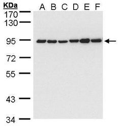
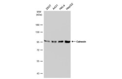
- Western blot analysis of Calnexin using 30 µg of A) 293T (B) A431 (C) H1299 (D) HeLa S3 (E) HepG2 and F) MOLT4 lysate. Samples were loaded onto a 7.5% SDS-PAGE gel and probed with a Calnexin polyclonal antibody (Product # PA5-34665) at a dilution of 1:5000.

- Submitted by
- Invitrogen Antibodies (provider)
- Main image
- Experimental details
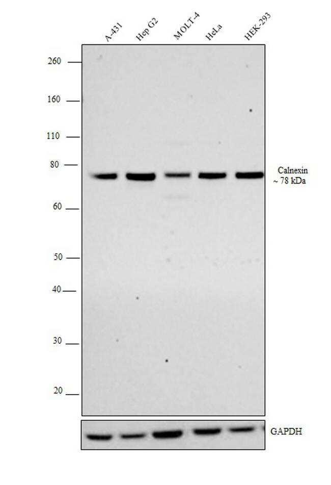
- Western blot analysis was performed on membrane enriched extracts (30 µg lysate) of A-431 (Lane 1), HepG2 (Lane 2), Molt-4 (Lane 3), HeLa (Lane 4) and HEK-293 (Lane 5). The blot was probed with Calnexin Polyclonal Antibody (Product # PA5-34665, 1:5000 dilution) and detected by chemiluminescence using Goat anti-Rabbit IgG (H+L) Superclonal™ Secondary Antibody, HRP conjugate (Product #, A27036, 0.25 µg/ml, 1:4000 dilution). A band at ~78 kDa corresponding to calnexin was observed across all the cell lines tested.
- Submitted by
- Invitrogen Antibodies (provider)
- Main image
- Experimental details
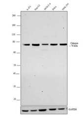
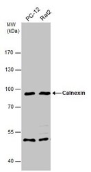
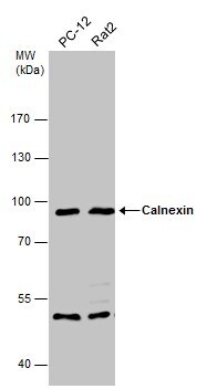
- Western Blot analysis of Calnexin was performed by separating 30 µg of various whole cell extracts by 7.5% SDS-PAGE. Proteins were transferred to a membrane and probed with a Calnexin Polyclonal Antibody (Product # PA5-34665) at a dilution of 1:5000 and a HRP-conjugated anti-rabbit IgG secondary antibody.

- Submitted by
- Invitrogen Antibodies (provider)
- Main image
- Experimental details
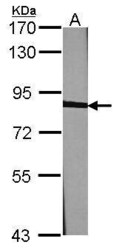
- Western Blot using Calnexin Polyclonal Antibody (Product # PA5-34665). Sample (30 µg of whole cell lysate). Lane A: NIH-3T3. 7.5% SDS PAGE. Calnexin Polyclonal Antibody (Product # PA5-34665) diluted at 1:1,000. The HRP-conjugated anti-rabbit IgG antibody was used to detect the primary antibody.
- Submitted by
- Invitrogen Antibodies (provider)
- Main image
- Experimental details
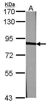
- Western Blot analysis of Calnexin was performed by separating 30 µg of various whole cell extracts by 7.5% SDS-PAGE. Proteins were transferred to a membrane and probed with a Calnexin Polyclonal Antibody (Product # PA5-34665) at a dilution of 1:1000 and a HRP-conjugated anti-rabbit IgG secondary antibody.
- Submitted by
- Invitrogen Antibodies (provider)
- Main image
- Experimental details
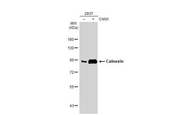
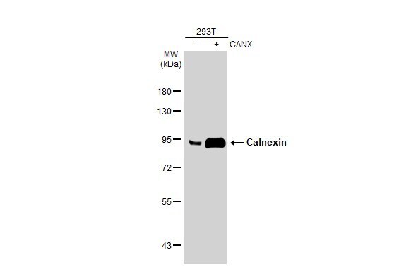
- Western Blot using Calnexin Polyclonal Antibody (Product # PA5-34665). Non-transfected (–) and transfected (+) 293T whole cell extracts (30 µg) were separated by 7.5% SDS-PAGE, and the membrane was blotted with Calnexin Polyclonal Antibody (Product # PA5-34665) diluted at 1:5,000. The HRP-conjugated anti-rabbit IgG antibody was used to detect the primary antibody.
- Submitted by
- Invitrogen Antibodies (provider)
- Main image

- Experimental details
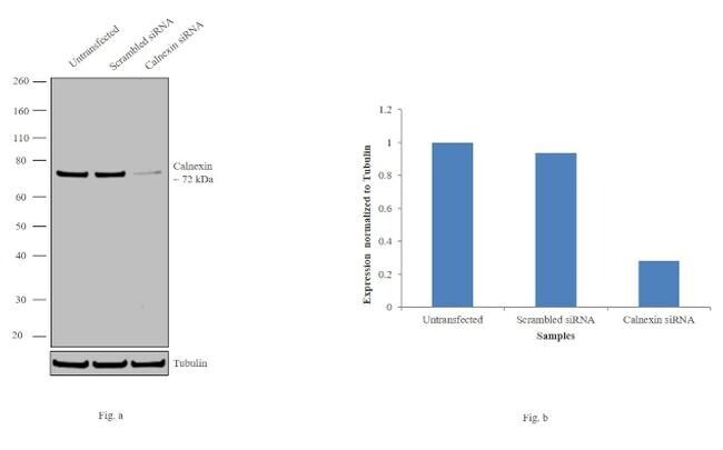
- Knockdown of Calnexin was achieved by transfecting HeLa cells with Calnexin specific siRNAs (Silencer® select Product # s2376). Western blot analysis (Fig. a) was performed using whole cell extracts from the Calnexin knockdown cells (lane 3), non-specific scrambled siRNA transfected cells (lane 2) and untransfected cells (lane 1). The blots were probed with Calnexin Polyclonal Antibody (Product # PA5-34665, 1:1000 dilution) and Goat anti-Rabbit IgG (H+L) Superclonal™ Secondary Antibody, HRP conjugate (Product # A27036, 0.25 µg/ml, 1:4000 dilution). Densitometric analysis of this western blot is shown in histogram (Fig. b). Decrease in signal upon siRNA mediated knock down confirms that antibody is specific to Calnexin.
Supportive validation
- Submitted by
- Invitrogen Antibodies (provider)
- Main image
- Experimental details
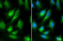
- Immunocytochemistry-Immunofluorescence analysis of Calnexin was performed in HeLa cells fixed in 4% paraformaldehyde at RT for 15 min. Green: Calnexin Polyclonal Antibody (Product # PA5-34665) diluted at 1:500. Blue: Hoechst 33342 staining. Scale bar = 10 µm.

Supportive validation
- Submitted by
- Invitrogen Antibodies (provider)
- Main image
- Experimental details
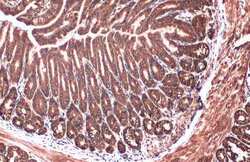
- Immunohistochemistry (Paraffin) analysis of Calnexin was performed in paraffin-embedded mouse duodenum tissue using Calnexin Polyclonal Antibody (Product # PA5-34665) at a dilution of 1:500. Antigen Retrieval: Citrate buffer, pH 6.0, 15 min.
- Submitted by
- Invitrogen Antibodies (provider)
- Main image

- Experimental details
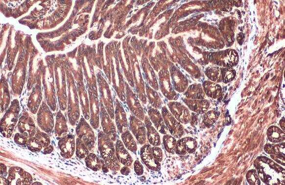
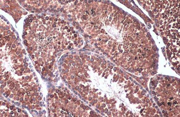
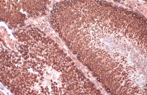
- Immunohistochemistry (Paraffin) analysis of Calnexin was performed in paraffin-embedded rat testis tissue using Calnexin Polyclonal Antibody (Product # PA5-34665) at a dilution of 1:500. Antigen Retrieval: Citrate buffer, pH 6.0, 15 min.
- Submitted by
- Invitrogen Antibodies (provider)
- Main image
- Experimental details
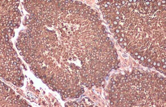
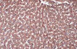
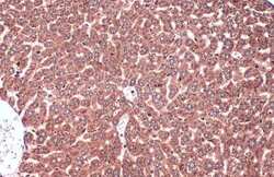
- Immunohistochemistry (Paraffin) analysis of Calnexin was performed in paraffin-embedded rat liver tissue using Calnexin Polyclonal Antibody (Product # PA5-34665) at a dilution of 1:500. Antigen Retrieval: Citrate buffer, pH 6.0, 15 min.
- Submitted by
- Invitrogen Antibodies (provider)
- Main image
- Experimental details
- Immunohistochemistry (Paraffin) analysis of Calnexin was performed in paraffin-embedded rat testis tissue using Calnexin Polyclonal Antibody (Product # PA5-34665) at a dilution of 1:500. Antigen Retrieval: Citrate buffer, pH 6.0, 15 min.
- Submitted by
- Invitrogen Antibodies (provider)
- Main image
- Experimental details
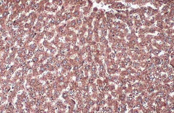
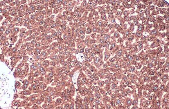
- Immunohistochemistry (Paraffin) analysis of Calnexin was performed in paraffin-embedded mouse liver tissue using Calnexin Polyclonal Antibody (Product # PA5-34665) at a dilution of 1:500. Antigen Retrieval: Citrate buffer, pH 6.0, 15 min.
- Submitted by
- Invitrogen Antibodies (provider)
- Main image

- Experimental details
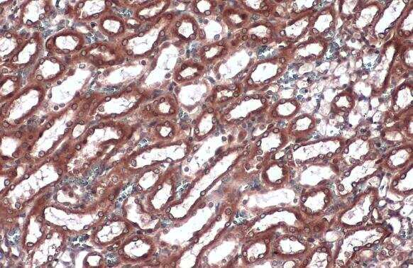
- Immunohistochemistry (Paraffin) analysis of Calnexin was performed in paraffin-embedded rat kidney tissue using Calnexin Polyclonal Antibody (Product # PA5-34665) at a dilution of 1:500. Antigen Retrieval: Citrate buffer, pH 6.0, 15 min.
- Submitted by
- Invitrogen Antibodies (provider)
- Main image
- Experimental details
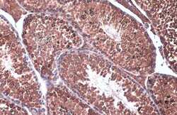
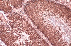
- Immunohistochemistry (Paraffin) analysis of Calnexin was performed in paraffin-embedded mouse testis tissue using Calnexin Polyclonal Antibody (Product # PA5-34665) at a dilution of 1:500. Antigen Retrieval: Citrate buffer, pH 6.0, 15 min.
Supportive validation
- Submitted by
- Invitrogen Antibodies (provider)
- Main image
- Experimental details
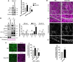
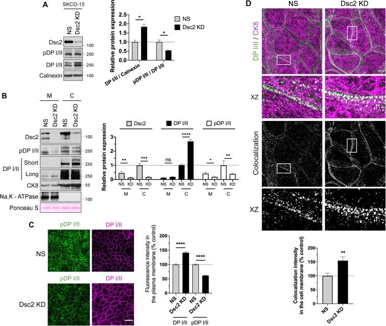
- FIGURE 7: Dsc2 KD resulted in reduced DP I/II phosphorylation at Ser2849 and enhanced interaction of DP I/II to IF-cytoskeleton. (A) Left panel shows representative WB images of the expression of Dsc2, pDP, DP I/II, and calnexin (loading control) in whole cell lysates derived from SKCO-15 Dsc2 KD vs. NS cells. Bar graphs in the right panel show densitometric analysis of DP I/II expression normalized to calnexin and ratio pDP/ DP I/II. Data represent mean +- SEM of three independent experiments. Statistical analysis was done with two-tailed Student's t test. * p < 0.05. (B) Membrane and cytoskeletal fractions were isolated from Dsc2 KD and control SKCO-15 cells. The left panel shows representative WB images of the expression of Dsc2, pDP I/II, DP I/II, Na,K - ATPase (membrane marker), and CK8-IF marker in Dsc2 KD vs. NS cells. Ponceau S staining serves as a loading control for total protein normalization. The right panel shows the densitometry analysis. Data represent mean +- SEM of five independent experiments. Statistical analysis was done with wo-tailed Student's t test. ns, not significant; * p < 0.05. ** p < 0.01; *** p < 0.001; **** p < 0.0001. (C) The left panel shows confocal microscopy images of pDP S2849 and DP I/II in SKCO-15 control (NS) vs. Dsc2 KD confluent monolayers. Scale bars are 40 mum. The right panel shows histograms depicting fluorescence intensity of DP I/II and pDP S2849 in the plasma membrane of KD confluent monolayers compared with NS control. Results
- Submitted by
- Invitrogen Antibodies (provider)
- Main image
- Experimental details
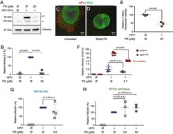
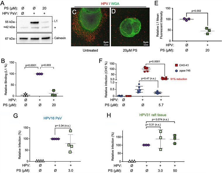
- FIG 3 Protamine interacts with cellular HSPGs to prevent HPV binding. (A) HaCaT cell monolayers were treated with 20 muM PS (in normal HaCaT medium) before the addition of HPV16 PsVs at 4degC for 1 h. After washing to remove unbound virus particles, cells were lysed and subjected to SDS-PAGE and immunoblot detection of HPV16 major capsid protein L1 (CAMVIR-1) and cellular calnexin as a loading control. A cellular protein at 42 kDa is nonspecifically (*) recognized by the CAMVIR-1 antibody (, ). (B) Densitometry quantification of HPV L1 normalized to calnexin ( n = 3 independent experiments) analyzed by one-way standard ANOVA. (C and D) Immunofluorescence performed on samples treated as in panel A. Cells were plated on coverslips and fixed with 4% PFA for immunofluorescence. HPV L1 (red) was stained using polyclonal anti-HPV16 VLP (1:200), and cell membranes were stained using the lectin wheat germ agglutinin (WGA; green). Representative images of untreated or PS-treated cells are shown. (E) ImageJ mean fluorescence intensity analysis of L1 signal. Each data point represents an average of three imaged areas from each independent experiment ( n = 3). (F) Parental CHO-K1 cells or HS- CHO cells (pgsa-745) were pretreated for 1 h with PS before being exposed to HPV16 PsVs. Infections were assessed by luciferase assay 24 h after PsV exposure ( n = 3 independent experiments). (G) HPV16 PsVs were allowed to interact with 3 muM PS (final) in a small volume for 1 h at 37degC prior
- Submitted by
- Invitrogen Antibodies (provider)
- Main image

- Experimental details
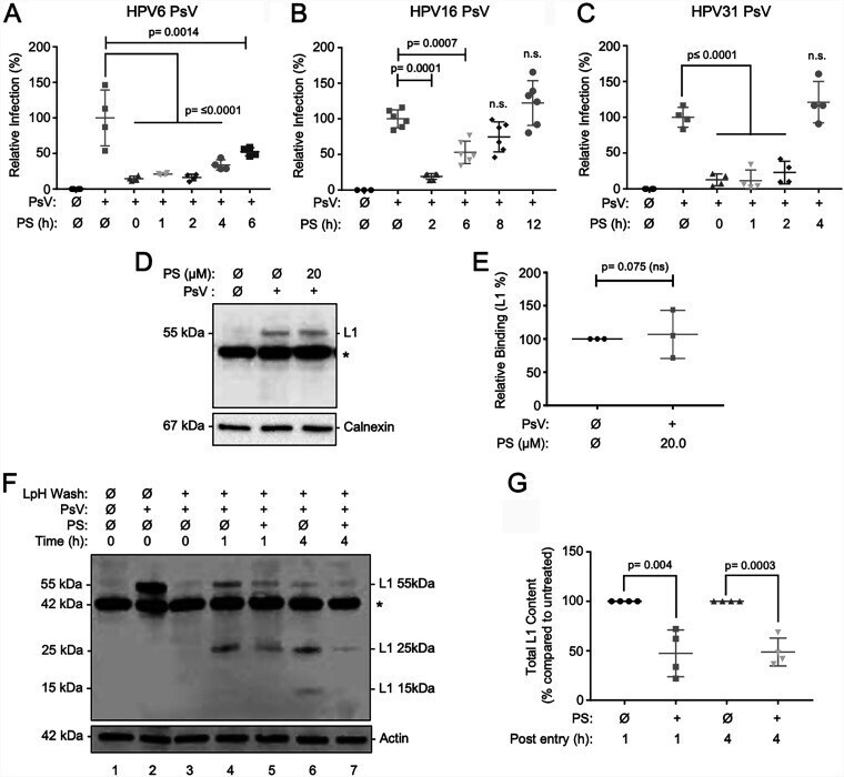
- FIG 4 Protamine reduces infection after viral attachment by slowing entry. (A to C) HaCaT cells were exposed to HPV PsV inocula for 1 h at 4degC to allow viral attachment. Inocula were removed, and the cells were washed to remove unbound virions. Thereafter, cells were treated with PS (5.7 muM final concentration) in complete medium at the indicated times after the transition to 37degC. Infections were allowed to proceed for a total of 24 h and assessed by luciferase assay. (A) HPV6 PsVs with 2 independent experiments each in duplicate. (B) HPV16 with 3 independent experiments each in duplicate. (C) HPV31 with 2 independent experiments each in duplicate analyzed by one-way standard ANOVA. P
- Submitted by
- Invitrogen Antibodies (provider)
- Main image

- Experimental details
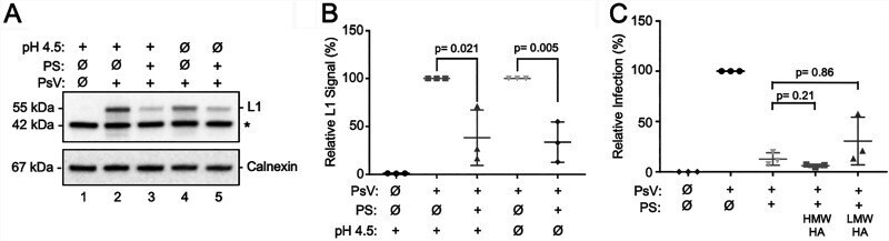
- FIG 6 Protamine sulfate reduces HPV binding under low-pH conditions and remains functional when mixed with hyaluronic acid (HA). (A) HaCaT cells were incubated in sodium lactate buffer (pH 4.5) containing 20 muM PS for 30 min prior to HPV16 PsV attachment at 4degC for 1 h. Unbound inocula were removed by washing; cellular lysates were collected and analyzed by SDS-PAGE and IB for HPV L1 and cellular calnexin as a loading control. *, nonspecific cellular reactant. (B) Densitometric quantification of L1 (55 kDa) normalized to calnexin ( n = 3 independent experiments) and analyzed by Student's t test. (C) HaCaT cells were treated with PS or PS incubated with high-molecular-weight (HMW; 1,500 to 1,800 kDa) HA or low-MW (LMW; 15 to 30 kDa) HA for 1 h at room temperature. PS-HA mixtures were used at final concentrations of 1 mg/mL HA and 3 muM PS. After treatment, cells were exposed to HPV16 PsVs, and infections were evaluated after 24 h by luciferase assay ( n = 3 independent experiments). Results were analyzed by one-way ANOVA.