Antibody data
- Antibody Data
- Antigen structure
- References [5]
- Comments [0]
- Validations
- Western blot [10]
- Immunocytochemistry [5]
- Immunohistochemistry [3]
- Other assay [5]
Submit
Validation data
Reference
Comment
Report error
- Product number
- PA5-34754 - Provider product page

- Provider
- Invitrogen Antibodies
- Product name
- Calnexin Polyclonal Antibody
- Antibody type
- Polyclonal
- Antigen
- Synthetic peptide
- Description
- Recommended positive controls: 293T, A431, HeLa, HepG2, A375, Neuro2A, GL261, C8D30, NIH-3T3, BCL-1, Raw264.7, C2C12, PC-12, Rat2, KN73. Predicted reactivity: Mouse (100%), Rat (100%), Dog (100%), Pig (100%), Bovine (100%). IHC notes, Requires antigen retrieval using heat mediated 10mM Citrate buffer (pH6.0) or Tris-EDTA buffer (pH8.0) Store product as a concentrated solution. Centrifuge briefly prior to opening the vial.
- Reactivity
- Human, Mouse, Rat
- Host
- Rabbit
- Isotype
- IgG
- Vial size
- 100 µL
- Concentration
- 0.46 mg/mL
- Storage
- Store at 4°C short term. For long term storage, store at -20°C, avoiding freeze/thaw cycles.
Submitted references Prevalence and mechanisms of evolutionary contingency in human influenza H3N2 neuraminidase.
Human Papillomavirus Minor Capsid Protein L2 Mediates Intracellular Trafficking into and Passage beyond the Endoplasmic Reticulum.
Striatal infusion of cholesterol promotes dose-dependent behavioral benefits and exerts disease-modifying effects in Huntington's disease mice.
Immunosuppression by Mutated Calreticulin Released from Malignant Cells.
Immunohistochemical staining reveals differential expression of ACSL3 and ACSL4 in hepatocellular carcinoma and hepatic gastrointestinal metastases.
Lei R, Tan TJC, Hernandez Garcia A, Wang Y, Diefenbacher M, Teo C, Gopan G, Tavakoli Dargani Z, Teo QW, Graham CS, Brooke CB, Nair SK, Wu NC
Nature communications 2022 Oct 28;13(1):6443
Nature communications 2022 Oct 28;13(1):6443
Human Papillomavirus Minor Capsid Protein L2 Mediates Intracellular Trafficking into and Passage beyond the Endoplasmic Reticulum.
Morante AV, Baboolal DD, Simon X, Pan EC, Meneses PI
Microbiology spectrum 2022 Jun 29;10(3):e0150522
Microbiology spectrum 2022 Jun 29;10(3):e0150522
Striatal infusion of cholesterol promotes dose-dependent behavioral benefits and exerts disease-modifying effects in Huntington's disease mice.
Birolini G, Valenza M, Di Paolo E, Vezzoli E, Talpo F, Maniezzi C, Caccia C, Leoni V, Taroni F, Bocchi VD, Conforti P, Sogne E, Petricca L, Cariulo C, Verani M, Caricasole A, Falqui A, Biella G, Cattaneo E
EMBO molecular medicine 2020 Oct 7;12(10):e12519
EMBO molecular medicine 2020 Oct 7;12(10):e12519
Immunosuppression by Mutated Calreticulin Released from Malignant Cells.
Liu P, Zhao L, Loos F, Marty C, Xie W, Martins I, Lachkar S, Qu B, Waeckel-Énée E, Plo I, Vainchenker W, Perez F, Rodriguez D, López-Otin C, van Endert P, Zitvogel L, Kepp O, Kroemer G
Molecular cell 2020 Feb 20;77(4):748-760.e9
Molecular cell 2020 Feb 20;77(4):748-760.e9
Immunohistochemical staining reveals differential expression of ACSL3 and ACSL4 in hepatocellular carcinoma and hepatic gastrointestinal metastases.
Ndiaye H, Liu JY, Hall A, Minogue S, Morgan MY, Waugh MG
Bioscience reports 2020 Apr 30;40(4)
Bioscience reports 2020 Apr 30;40(4)
No comments: Submit comment
Supportive validation
- Submitted by
- Invitrogen Antibodies (provider)
- Main image

- Experimental details
- Western blot analysis of Calnexin using Various whole cell extracts (30 µg). Samples were loaded onto a 7.5% SDS-PAGE gel and probed with a Calnexin polyclonal antibody (Product # PA5-34754) at a dilution of 1:10,000.
- Submitted by
- Invitrogen Antibodies (provider)
- Main image

- Experimental details
- Western blot analysis of Calnexin using 30 µg of A) NIH-3T3 (B) JC and C) BCL-1 lysate. Samples were loaded onto a 7.5% SDS-PAGE gel and probed with a Calnexin polyclonal antibody (Product # PA5-34754) at a dilution of 1:1000.
- Submitted by
- Invitrogen Antibodies (provider)
- Main image

- Experimental details
- Western Blot analysis of Calnexin was performed by separating 30 µg of various whole cell extracts by 7.5% SDS-PAGE. Proteins were transferred to a membrane and probed with a Calnexin Polyclonal Antibody (Product # PA5-34754) at a dilution of 1:10000 and a HRP-conjugated anti-rabbit IgG secondary antibody.
- Submitted by
- Invitrogen Antibodies (provider)
- Main image

- Experimental details
- Western blot analysis was performed on whole cell extracts (30 µg lysate) of HeLa (Lane 1), HCT116 (Lane 2), A431 (Lane 3), HepG2 (Lane 4), Raji (Lane 5), MOLT-4 (Lane 6), Jurkat (Lane 7) and RAW 264.7 (Lane 8). The blot was probed with Anti- Calnexin Polyclonal Antibody (Product # PA5-34754, 1:5000 dilution) and detected by chemiluminescence using Goat anti-Rabbit IgG (H+L) Superclonal™ Secondary Antibody, HRP conjugate (Product # A27036, 0.25 µg/ml, 1:4000 dilution). A 72 kDa band corresponding to Calnexin was observed across all the cell lines tested.
- Submitted by
- Invitrogen Antibodies (provider)
- Main image

- Experimental details
- Western Blot analysis of Calnexin was performed by separating 30 µg of various whole cell extracts by 7.5% SDS-PAGE. Proteins were transferred to a membrane and probed with a Calnexin Polyclonal Antibody (Product # PA5-34754) at a dilution of 1:10000 and a HRP-conjugated anti-rabbit IgG secondary antibody.
- Submitted by
- Invitrogen Antibodies (provider)
- Main image

- Experimental details
- Western Blot analysis of Calnexin was performed by separating 30 µg of various whole cell extracts by 7.5% SDS-PAGE. Proteins were transferred to a membrane and probed with a Calnexin Polyclonal Antibody (Product # PA5-34754) at a dilution of 1:10000 and a HRP-conjugated anti-rabbit IgG secondary antibody.
- Submitted by
- Invitrogen Antibodies (provider)
- Main image

- Experimental details
- Western Blot using Calnexin Polyclonal Antibody (Product # PA5-34754). Various whole cell extracts (30 µg) were separated by 7.5% SDS-PAGE, and the membrane was blotted with Calnexin Polyclonal Antibody (Product # PA5-34754) diluted at 1:10,000. The HRP-conjugated anti-rabbit IgG antibody was used to detect the primary antibody.
- Submitted by
- Invitrogen Antibodies (provider)
- Main image

- Experimental details
- Western Blot analysis of Calnexin was performed by separating 30 µg of various whole cell lysates by 7.5 % SDS-PAGE. Proteins were transferred to a membrane and probed with a Calnexin Polyclonal Antibody (Product # PA5-34754) at a dilution of 1:10000. A. Neuro2A, B. GL261, C. C8D30, D. NIH-3T3, E. BCL-1, F. Raw264.7, G. C2C12.
- Submitted by
- Invitrogen Antibodies (provider)
- Main image

- Experimental details
- Western Blot using Calnexin Polyclonal Antibody (Product # PA5-34754). Non-transfected (–) and transfected (+) 293T whole cell extracts (30 µg) were separated by 7.5% SDS-PAGE, and the membrane was blotted with Calnexin Polyclonal Antibody (Product # PA5-34754) diluted at 1:5,000. The HRP-conjugated anti-rabbit IgG antibody was used to detect the primary antibody.
- Submitted by
- Invitrogen Antibodies (provider)
- Main image

- Experimental details
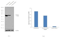
- Knockdown of Calnexin was achieved by transfecting HeLa cells with Calnexin specific siRNAs (Silencer® select Product # s2376). Western blot analysis (Fig. a) was performed using whole cell extracts from the Calnexin knockdown cells (lane 3), non-specific scrambled siRNA transfected cells (lane 2) and untransfected cells (lane 1). The blot was probed with Calnexin Polyclonal Antibody (Product # PA5-34754, 1:2000 dilution) and Goat anti-Rabbit IgG (H+L) Superclonal™ Secondary Antibody, HRP conjugate (Product # A27036, 0.25µg/ml, 1:4000 dilution). Densitometric analysis of this western blot is shown in histogram (Fig. b). Decrease in signal upon siRNA mediated knock down confirms that antibody is specific to Calnexin.
Supportive validation
- Submitted by
- Invitrogen Antibodies (provider)
- Main image

- Experimental details
- Immunofluorescent analysis of Calnexin in paraformaldehyde-fixed HeLa cells using a Calnexin polyclonal antibody (Product # PA5-34754) (Green) at a 1:200 dilution. Alpha-tubulin filaments were labeled with Product # PA5-29281 (Red) at a 1:2500.
- Submitted by
- Invitrogen Antibodies (provider)
- Main image

- Experimental details
- Immunofluorescent analysis of Calnexin in methanol-fixed HeLa cells using a Calnexin polyclonal antibody (Product # PA5-34754) (Green) at a 1:500 dilution. Alpha-tubulin filaments were labeled with Product # PA5-29281 (Red) at a 1:2500.
- Submitted by
- Invitrogen Antibodies (provider)
- Main image

- Experimental details
- Immunofluorescence analysis of Calnexin was performed using 70% confluent log phase HeLa cells treated with Thapsigargin (1uM for 24hrs). The cells were fixed with 4% paraformaldehyde for 10 minutes, permeabilized with 0.1% Triton™ X-100 for 15 minutes, and blocked with 1% BSA for 1 hour at room temperature. The cells were labeled with Calnexin Polyclonal Antibody (Product # PA5-34754) at 1:200 dilution in 0.1% BSA, incubated at 4 degree celsius overnight and then labeled with Goat anti-Rabbit IgG (H+L) Superclonal™ Secondary Antibody, Alexa Fluor® 594 conjugate (Product # A-11062) at a dilution of 1:2000 for 45 minutes at room temperature (Panel a: red). Nuclei (Panel b: blue) were stained with ProLong™ Diamond Antifade Mountant with DAPI (Product # P36962). F-actin (Panel c: green) was stained with Alexa Fluor™ 488 Phalloidin (Product # A12379, 1:300). Panel d represents the merged image showing Calnexin in the ER and cytoplasm. Panel e represents the untreated cells showing lower expression levels. Panel f represents control cells with no primary antibody to assess background. The images were captured at 60X magnification.
- Submitted by
- Invitrogen Antibodies (provider)
- Main image

- Experimental details
- Immunocytochemistry-Immunofluorescence analysis of Calnexin was performed in DIV9 rat E18 primary cortical neuron cells fixed in 4% paraformaldehyde at RT for 15 min. Green: Calnexin Polyclonal Antibody (Product # PA5-34754) diluted at 1:500. Red: beta Tubulin 3/ Tuj1, stained by beta Tubulin 3/ Tuj1 antibody. Blue: Fluoroshield with DAPI.
- Submitted by
- Invitrogen Antibodies (provider)
- Main image

- Experimental details
- Calnexin Polyclonal Antibody detects Calnexin protein at endoplasmic reticulum by immunofluorescent analysis. Sample: HeLa cells were fixed in 4% paraformaldehyde at RT for 15 min. Green: Calnexin stained by Calnexin Polyclonal Antibody (Product # PA5-34754) diluted at 1:500. Blue: Fluoroshield with DAPI . Scale bar= 10 µm.
Supportive validation
- Submitted by
- Invitrogen Antibodies (provider)
- Main image

- Experimental details
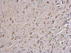
- Calnexin Polyclonal Antibody detects Calnexin protein at cytosol on mouse middle brain by immunohistochemical analysis. Sample: Paraffin-embedded mouse middle brain. Calnexin Polyclonal Antibody (Product # PA5-34754) dilution: 1:500. Antigen Retrieval: EDTA based buffer, pH 8.0, 15 min.
- Submitted by
- Invitrogen Antibodies (provider)
- Main image

- Experimental details
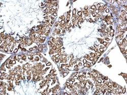
- Calnexin Polyclonal Antibody detects Calnexin protein at cytosol on mouse testis by immunohistochemical analysis. Sample: Paraffin-embedded mouse testis. Calnexin Polyclonal Antibody (Product # PA5-34754) dilution: 1:500. Antigen Retrieval: EDTA based buffer, pH 8.0, 15 min.
- Submitted by
- Invitrogen Antibodies (provider)
- Main image

- Experimental details
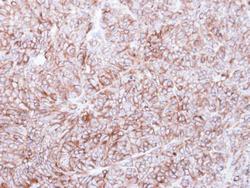
- Immunohistochemical analysis of paraffin-embedded DLD-1 xenograft, using Calnexin (Product # PA5-34754) antibody at 1:500 dilution. Antigen Retrieval: EDTA based buffer, pH 8.0, 15 min.
Supportive validation
- Submitted by
- Invitrogen Antibodies (provider)
- Main image

- Experimental details
- Calnexin antibody immunoprecipitates Calnexin protein in IP experiments. IP Sample: 1,000 µg HeLa whole cell lysate/extract A. 30 µg HeLa whole cell lysate/extract B. Control with 2 µg of preimmune rabbit IgG C. Immunoprecipitation of Calnexin protein by 2 µg of Calnexin antibody (Product # PA5-34754) 7.5% SDS-PAGE The immunoprecipitated Calnexin protein was detected by Calnexin antibody (Product # PA5-34754) diluted at 1:1,000.
- Submitted by
- Invitrogen Antibodies (provider)
- Main image

- Experimental details
- EV1 Behavioral characterization of wt mice following striatal infusion of the high dose of cholesterol and in vivo exogenous cholesterol localization A Latency to fall (seconds) from an accelerating rotarod at 10 weeks of age (3 weeks after cholesterol infusion) in wt ( N = 11); wt ACSF ( N = 11) and wt chol-high ( N = 7) mice. B-E Global motor activity (B), total distance travelled (C), mean velocity (D), and stereotyped movements (E) in an open field at 11 weeks of age (4 weeks after cholesterol infusion) (wt = 11; wt ACSF = 10; wt chol-high = 7). F Discrimination index (%) in the novel object recognition test of wt, wt ACSF, and wt chol-high mice at 11 weeks of age (4 weeks after cholesterol infusion) (wt = 11; wt ACSF = 10; wt chol-high = 7). DI above zero indicates a preference for the novel object; DI below zero indicates a preference for the familiar object. G, H Cholesterol content in the infused striatum (G) and ipsilateral cortex (H) of wt ACSF ( N = 3), wt chol-low ( N = 5), and wt chol-high ( N = 4) mice at 12 weeks of age after 4-week striatal cholesterol infusion. I-M Representative confocal images showing co-localization of BODIPY-chol (green) and TGN46 (I), calnexin (J), Rab9 (K), PMCA-ATPase (L), and LAMP1 (M) (red) in the striatum of R6/2 mice infused with BODIPY-cholesterol. Scale bars: 5 mum. Data information: The data in (A-H) are shown as scatterplot graphs with means +- standard error. Each dot corresponds to the value obtained from each animal. Values
- Submitted by
- Invitrogen Antibodies (provider)
- Main image

- Experimental details
- Figure 6 Equilibrium distributions of ACSL3 and ACSL4 in sucrose density gradient fractions prepared from HepG2 cells Subcellular fractions isolated from HepG2 cells were separated by SDS/PAGE and Western blots were carried out to detect the lipid-droplet protein PNPLA3, ACSL3 and ACSL4, the ER marker protein calnexin, plasma membrane and lipid-raft associated flotillin, the Golgi protein GS28, the TGN-endosomal protein syntaxin-6, the early endosome-recruited protein EEA and the mitochondrial protein VDAC. Western blots are representative of experiments repeated three to four times.
- Submitted by
- Invitrogen Antibodies (provider)
- Main image

- Experimental details
- Figure EV1 Behavioral characterization of wt mice following striatal infusion of the high dose of cholesterol and in vivo exogenous cholesterol localization A Latency to fall (seconds) from an accelerating rotarod at 10 weeks of age (3 weeks after cholesterol infusion) in wt ( N = 11); wt ACSF ( N = 11) and wt chol-high ( N = 7) mice. B-E Global motor activity (B), total distance travelled (C), mean velocity (D), and stereotyped movements (E) in an open field at 11 weeks of age (4 weeks after cholesterol infusion) (wt = 11; wt ACSF = 10; wt chol-high = 7). F Discrimination index (%) in the novel object recognition test of wt, wt ACSF, and wt chol-high mice at 11 weeks of age (4 weeks after cholesterol infusion) (wt = 11; wt ACSF = 10; wt chol-high = 7). DI above zero indicates a preference for the novel object; DI below zero indicates a preference for the familiar object. G, H Cholesterol content in the infused striatum (G) and ipsilateral cortex (H) of wt ACSF ( N = 3), wt chol-low ( N = 5), and wt chol-high ( N = 4) mice at 12 weeks of age after 4-week striatal cholesterol infusion. I-M Representative confocal images showing co-localization of BODIPY-chol (green) and TGN46 (I), calnexin (J), Rab9 (K), PMCA-ATPase (L), and LAMP1 (M) (red) in the striatum of R6/2 mice infused with BODIPY-cholesterol. Scale bars: 5 mum. Data information: The data in (A-H) are shown as scatterplot graphs with means +- standard error. Each dot corresponds to the value obtained from each animal.
- Submitted by
- Invitrogen Antibodies (provider)
- Main image

- Experimental details
- FIG 7 HPV16 pseudovirions associate with the endoplasmic reticulum 20 hpi. Cells were seeded onto coverslips and infected with WT, DeltaQ mutant, or DeltaIL mutant PsVs for 20 h before immunofluorescence slide preparation and antibody staining for either EdU and calnexin overlap (A-L) or H16.V5 and calnexin overlap (M-X) Representative immunofluorescence images are taken from the center of Z-stack image set. Top three panels represent EdU and calnexin signal for WT (A-D), DeltaQ mutant (E-H), and DeltaIL mutant (I-L) PsVs. (A, E, I) Nuclei stained with DAPI (gray). (B, F, J) Pseudogenome expressing EdU (red). (C, G, K) ER stained with calnexin (green). (D, H, L) Merged image of all channels displaying colocalization of pseudogenome and calnexin. (D1, H1, L1) Zoomed images of white square from merged channels. Bottom three panels represent H16.V5 and calnexin signal for WT (M-P), DeltaQ mutant (Q-T), and DeltaIL mutant (U-X) PsVs. (M, Q, U) Nuclei stained with DAPI (gray). (N, R, V) H16.V5 representing HPV16 L1 protein (red). (O, S, W) ER stained with calnexin (green). (P, T, X) Merged image of all channels displaying colocalization of H16.V5 and calnexin. (P1, T1, X1) Zoomed images of white square from merged channels. (Y-Z) M1 coefficient for (Y) pseudogenome (EdU) or (Z) H16.V5 overlapping ER membrane (calnexin) at 20 h postinfection for untreated, Optiprep, O plasmid preparation, WT PsVs, DeltaQ mutant PsVs, and DeltaIL mutant PsVs. Data is the average of six independent c
 Explore
 Validate
 Learn
Learn Western blot
 Immunoprecipitation