Explore
 Validate
 Learn
Learn Western blot
Antibody data
- Antibody Data
- Antigen structure
- References [0]
- Comments [0]
- Validations
- Western blot [3]
- Immunocytochemistry [1]
- Flow cytometry [1]
Submit
Validation data
Reference
Comment
Report error
- Product number
- F44713 - Provider product page

- Provider
- NSJ Bioreagents
- Product name
- KRAS Antibody
- Antibody type
- Polyclonal
- Antigen
- A portion of amino acids 146-174 from the human protein was used as the immunogen for this KRAS antibody.
- Description
- Antigen affinity purified antibody
- Reactivity
- Human, Mouse
- Host
- Rabbit
- Conjugate
- Unconjugated
- Vial size
- 80 µl, 400 µl
- Storage
- Aliquot the KRAS antibody and store frozen at -20°C or colder. Avoid repeated freeze-thaw cycles.
No comments: Submit comment
Supportive validation
- Submitted by
- NSJ Bioreagents (provider)
- Main image
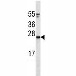
- Experimental details
- KRAS antibody western blot analysis in mouse NIH3T3 lysate. Predicted molecular weight: 20-25 kDa

- Submitted by
- NSJ Bioreagents (provider)
- Main image
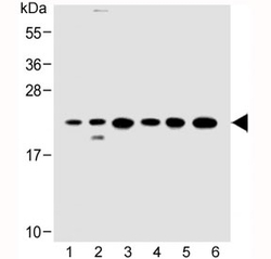
- Experimental details
- Western blot testing of 1) human HeLa, 2) human 293/T17, 3) mouse C2C12, 4) rat C6, 5) human HT-29 and 6) rat PC-12 cell lysate with KRAS antibody at 1:500. Expected molecular weight: 20-25 kDa.

- Submitted by
- NSJ Bioreagents (provider)
- Main image
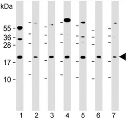
- Experimental details
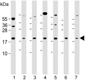
- Western blot testing of 1) human 293T/17, 2) mouse C2C12, 3) human HeLa, 4) human HT-29, 5) human K562, 6) rat PC-12 and 7) human Ramos cell lysate with KRAS antibody at 1:2000. Expected molecular weight: 20-25 kDa.
Supportive validation
- Submitted by
- NSJ Bioreagents (provider)
- Main image
- Experimental details
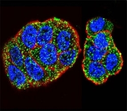
- Confocal immunofluorescent analysis of KRAS antibody with WiDr cells followed by Alexa Fluor 488-conjugated goat anti-rabbit lgG (green). Actin filaments have been labeled with Alexa Fluor 555 Phalloidin (red). DAPI was used as a nuclear counterstain (blue).

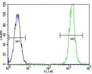
Supportive validation
- Submitted by
- NSJ Bioreagents (provider)
- Main image
- Experimental details
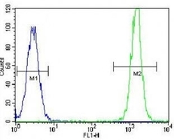
- KRAS antibody flow cytometric analysis of HeLa cells (right histogram) compared to a negative control (left histogram). FITC-conjugated goat-anti-rabbit secondary Ab was used for the analysis.