Explore
 Validate
 Learn
Learn Western blot
Antibody data
- Antibody Data
- Antigen structure
- References [0]
- Comments [0]
- Validations
- Western blot [3]
- Immunohistochemistry [3]
Submit
Validation data
Reference
Comment
Report error
- Product number
- PA5-73003 - Provider product page

- Provider
- Invitrogen Antibodies
- Product name
- OLFM4 Polyclonal Antibody
- Antibody type
- Polyclonal
- Antigen
- Synthetic peptide
- Reactivity
- Human, Mouse
- Host
- Rabbit
- Isotype
- IgG
- Vial size
- 100 µg
- Concentration
- 1.0 mg/mL
- Storage
- -20° C, Avoid Freeze/Thaw Cycles
No comments: Submit comment
Supportive validation
- Submitted by
- Invitrogen Antibodies (provider)
- Main image
- Experimental details
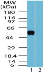
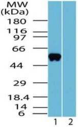
- Western blot analysis of OLFM4 in human liver (1: no immunizing peptide; 2: with immunizing peptide) tissue lysate using OLFM4 polyclonal antibody (Product # PA5-73003) at a dilution on 2 µg/mL.

- Submitted by
- Invitrogen Antibodies (provider)
- Main image
- Experimental details
- Western blot analysis of OLFM4 in human liver lysate in the 1) absence and 2) presence of immunizing peptide. Samples were incubated in OLFM4 polyclonal antibody (Product # PA5-73003) using a dilution of 2 µg/mL.

- Submitted by
- Invitrogen Antibodies (provider)
- Main image
- Experimental details
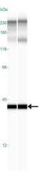
- Western blot analysis of OLFM4 in 0.2 mg/mL OLFM4. Samples were incubated in OLFM4 polyclonal antibody (Product # PA5-73003). This experiment was performed under reducing conditions using the 12-230 kDa separation system. *Non-specific interaction with the 230 kDa standard may be seen with this antibody.

Supportive validation
- Submitted by
- Invitrogen Antibodies (provider)
- Main image
- Experimental details
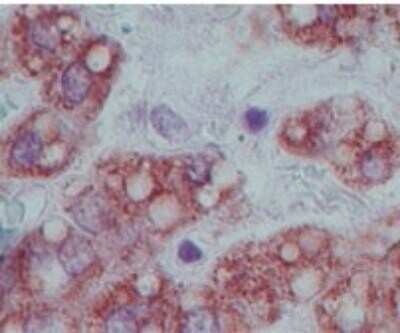
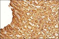
- Immunohistochemical analysis of OLFM4 in Tissue section of mouse stomach. Samples were incubated in OLFM4 polyclonal antibody (Product # PA5-73003) using a dilution of 2.5 µg/mL followed by HRP-DAB detection and hematoxylin counterstaining. The antibody generated very specific staining for OLFM4 in almost all cell types. The signal was localized specifically to the inter-cellular spaces, the nuclei and the cytoplasm. The simple columnar epithelial cells showed stronger and a distinct inter-cellular signal compared to the cells of the gastric glands.

- Submitted by
- Invitrogen Antibodies (provider)
- Main image
- Experimental details
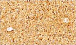
- Immunohistochemical analysis of OLFM4 in Tissue section of mouse liver. Samples were incubated in OLFM4 polyclonal antibody (Product # PA5-73003) using a dilution of 2.5 µg/mL followed by HRP-DAB detection and hematoxylin counterstaining. The antibody generated nice staining in all the hepatocytes and the signal was localized specifically to the inter-cellular spaces, the nuclei and the cytoplasm.
- Submitted by
- Invitrogen Antibodies (provider)
- Main image
- Experimental details
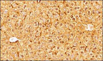
- Immunohistochemical analysis of OLFM4 in formalin-fixed, paraffin-embedded human liver tissue. Samples were incubated in OLFM4 polyclonal antibody (Product # PA5-73003) using a dilution of 5 µg/mL.