 Explore
 Validate
 Learn
Learn Western blot
 Immunocytochemistry
Antibody data
- Antibody Data
- Antigen structure
- References [2]
- Comments [0]
- Validations
- Western blot [1]
- Immunohistochemistry [1]
- Flow cytometry [2]
Submit
Validation data
Reference
Comment
Report error
- Product number
- MAB7367 - Provider product page

- Provider
- Novus Biologicals
- Product name
- Rat Monoclonal Desmocollin-1 Antibody
- Antibody type
- Monoclonal
- Description
- Protein A or G purified from hybridoma culture supernatant. Detects mouse Desmocollin-1 in ELISAs and Western blots. In direct ELISAs, 100% cross-reactivity with recombinant human Desmocollin-1 is observed, and no cross-reactivity with recombinant mouse Desmocollin-2 or -3 is observed.
- Reactivity
- Human, Mouse
- Host
- Rat
- Conjugate
- Unconjugated
- Isotype
- IgG
- Vial size
- 100 ug
- Concentration
- LYOPH
- Storage
- Use a manual defrost freezer and avoid repeated freeze-thaw cycles. 12 months from date of receipt, -20 to -70 degreesC as supplied. 1 month, 2 to 8 degreesC under sterile conditions after reconstitution. 6 months, -20 to -70 degreesC under sterile conditions after reconstitution.
Submitted references BubR1 Insufficiency Impairs Liver Regeneration in Aged Mice after Hepatectomy through Intercalated Disc Abnormality.
A spontaneous deletion within the desmoglein 3 extracellular domain of mice results in hypomorphic protein expression, immunodeficiency, and a wasting disease phenotype.
Ikawa-Yoshida A, Matsumoto T, Okano S, Aoyagi Y, Matsubara Y, Furuyama T, Nakatsu Y, Tsuzuki T, Onimaru M, Ohkusa T, Nomura M, Maehara Y
Scientific reports 2016 Aug 26;6:32399
Scientific reports 2016 Aug 26;6:32399
A spontaneous deletion within the desmoglein 3 extracellular domain of mice results in hypomorphic protein expression, immunodeficiency, and a wasting disease phenotype.
Kountikov EI, Poe JC, Maclver NJ, Rathmell JC, Tedder TF
The American journal of pathology 2015 Mar;185(3):617-30
The American journal of pathology 2015 Mar;185(3):617-30
No comments: Submit comment
Supportive validation
- Submitted by
- Novus Biologicals (provider)
- Main image

- Experimental details
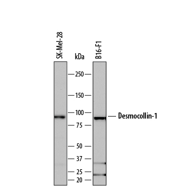
- Detection of Human and Mouse Desmocollin-1 by Western Blot. Western blot shows lysates of SK-Mel-28 human malignant melanoma cell line and B16-F1 mouse melanoma cell line. PVDF membrane was probed with 0.1 µg/mL of Rat Anti-Human/Mouse Desmocollin-1 Monoclonal Antibody (Catalog # MAB7367) followed by HRP-conjugated Anti-Rat IgG Secondary Antibody (Catalog # HAF005). A specific band was detected for Desmocollin-1 at approximately 98 kDa (as indicated). This experiment was conducted under reducing conditions and using Immunoblot Buffer Group 1.
Supportive validation
- Submitted by
- Novus Biologicals (provider)
- Main image

- Experimental details
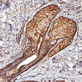
- Desmocollin-1 in Mouse Skin. Desmocollin-1 was detected in perfusion fixed frozen sections of mouse skin using Rat Anti-Human/Mouse Desmocollin-1 Monoclonal Antibody (Catalog # MAB7367) at 1.7 µg/mL overnight at 4 °C. Tissue was stained using the Anti-Rat HRP-DAB Cell & Tissue Staining Kit (brown; Catalog # CTS017) and counterstained with hematoxylin (blue). Specific staining was localized to hair root sheath. View our protocol for Chromogenic IHC Staining of Frozen Tissue Sections.
Supportive validation
- Submitted by
- Novus Biologicals (provider)
- Main image

- Experimental details
- Detection of Desmocollin-1 in A549 Human Cell Line by Flow Cytometry. A549 human lung carcinoma cell line was stained with Rat Anti-Human/Mouse Desmocollin-1 Monoclonal Antibody (Catalog # MAB7367, filled histogram) or isotype control antibody (Catalog # MAB006, open histogram), followed by Allophycocyanin-conjugated Anti-Rat IgG Secondary Antibody (Catalog # F0113).

- Submitted by
- Novus Biologicals (provider)
- Main image

- Experimental details
- Detection of Desmocollin-1 in B16-F1 Mouse Cell Line by Flow Cytometry. B16-F1 mouse melanoma cell line was stained with Rat Anti-Human/Mouse Desmocollin-1 Monoclonal Antibody (Catalog # MAB7367, filled histogram) or isotype control antibody (Catalog # MAB006, open histogram), followed by Allophycocyanin-conjugated Anti-Rat IgG Secondary Antibody (Catalog # F0113).
