Explore
 Validate
 Learn
Learn Western blot
Antibody data
- Antibody Data
- Antigen structure
- References [0]
- Comments [0]
- Validations
- Western blot [4]
- Immunocytochemistry [1]
- Immunohistochemistry [9]
- Flow cytometry [1]
Submit
Validation data
Reference
Comment
Report error
- Product number
- PA5-79856 - Provider product page

- Provider
- Invitrogen Antibodies
- Product name
- Periplakin Polyclonal Antibody
- Antibody type
- Polyclonal
- Antigen
- Synthetic peptide
- Description
- Reconstitute with 0.2 mL of distilled water to yield a concentration of 500 µg/mL.
- Reactivity
- Human, Mouse, Rat
- Host
- Rabbit
- Isotype
- IgG
- Vial size
- 100 µg
- Concentration
- 500 µg/mL
- Storage
- -20°C
No comments: Submit comment
Supportive validation
- Submitted by
- Invitrogen Antibodies (provider)
- Main image

- Experimental details
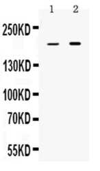
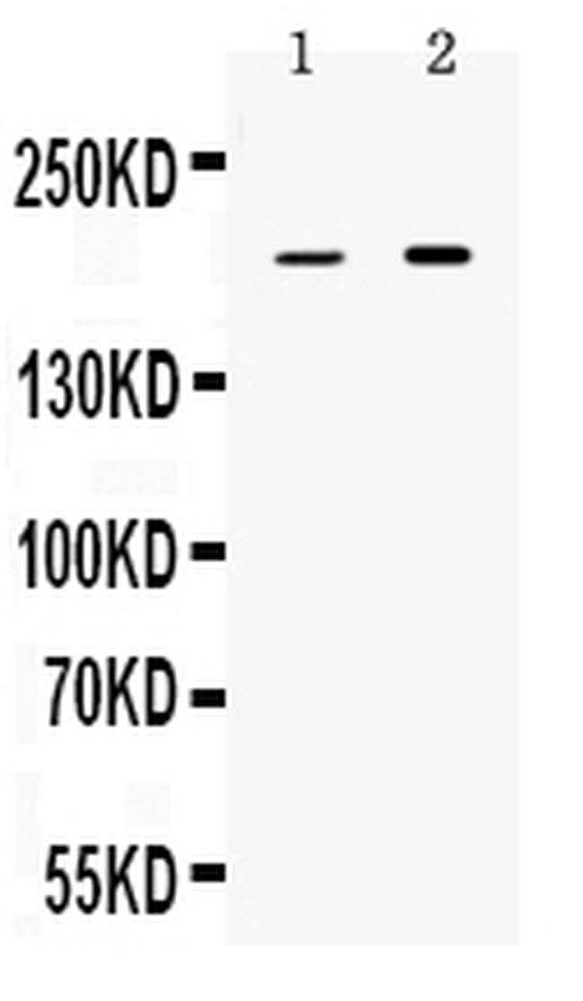
- Western blot analysis of Periplakin in rat stomach extract (lane 1) and HeLa whole cell lysate (lane 2). Sample was incubated with Periplakin polyclonal antibody (Product # PA5-79856) at a dilution of 0.5 µg/mL. Signal development was performed using a chemiluminescence (ECL) kit.

- Submitted by
- Invitrogen Antibodies (provider)
- Main image
- Experimental details
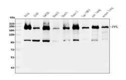
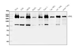
- Western blot analysis of Periplakin using anti-Periplakin antibody (Product # PA5-79856). Electrophoresis was performed on a 5-20% SDS-PAGE gel at 70V (Stacking gel)/90V (Resolving gel) for 2-3 hours. The sample well of each lane was loaded with 30 µg of sample under reducing conditions. Lane 1: human Hela whole cell lysates, Lane 2: human A549 whole cell lysates, Lane 3: human SW620 whole cell lysates, Lane 4: human HepG2 whole cell lysates, Lane 5: human SK-OV-3 whole cell lysates, Lane 6: human PANC-1 whole cell lysates, Lane 7: human SGC-7801 whole cell lysates, Lane 8: rat lung tissue lysates, Lane 9: mouse lung tissue lysates. After electrophoresis, proteins were transferred to a nitrocellulose membrane at 150 mA for 50-90 minutes. Membrane was blocked with 5% non-fat milk/TBS for 1.5 hour at RT. The membrane was incubated with rabbit anti-Periplakin antigen affinity purified polyclonal antibody (Product # PA5-79856) at 0.25 µg/mL overnight at 4°C and probed with a goat anti-rabbit IgG-HRP secondary antibody at a dilution of 1:5,000 for 1.5 hour at RT. The signal is developed using an Enhanced Chemiluminescent detection (ECL) kit. A specific band was detected at approximately 202 kDa. The expected band size is at 205 kDa.
- Submitted by
- Invitrogen Antibodies (provider)
- Main image
- Experimental details
- Western blot analysis of Periplakin in rat stomach extract (lane 1) and HeLa whole cell lysate (lane 2). Sample was incubated with Periplakin polyclonal antibody (Product # PA5-79856) at a dilution of 0.5 µg/mL. Signal development was performed using a chemiluminescence (ECL) kit.
- Submitted by
- Invitrogen Antibodies (provider)
- Main image
- Experimental details
- Western blot analysis of Periplakin using anti-Periplakin antibody (Product # PA5-79856). Electrophoresis was performed on a 5-20% SDS-PAGE gel at 70V (Stacking gel)/90V (Resolving gel) for 2-3 hours. The sample well of each lane was loaded with 30 µg of sample under reducing conditions. Lane 1: human Hela whole cell lysates, Lane 2: human A549 whole cell lysates, Lane 3: human SW620 whole cell lysates, Lane 4: human HepG2 whole cell lysates, Lane 5: human SK-OV-3 whole cell lysates, Lane 6: human PANC-1 whole cell lysates, Lane 7: human SGC-7801 whole cell lysates, Lane 8: rat lung tissue lysates, Lane 9: mouse lung tissue lysates. After electrophoresis, proteins were transferred to a nitrocellulose membrane at 150 mA for 50-90 minutes. Membrane was blocked with 5% non-fat milk/TBS for 1.5 hour at RT. The membrane was incubated with rabbit anti-Periplakin antigen affinity purified polyclonal antibody (Product # PA5-79856) at 0.25 µg/mL overnight at 4°C and probed with a goat anti-rabbit IgG-HRP secondary antibody at a dilution of 1:5,000 for 1.5 hour at RT. The signal is developed using an Enhanced Chemiluminescent detection (ECL) kit. A specific band was detected at approximately 202 kDa. The expected band size is at 205 kDa.

Supportive validation
- Submitted by
- Invitrogen Antibodies (provider)
- Main image
- Experimental details
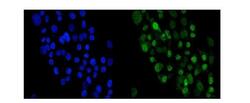
- Immunocytochemical analysis of Periplakin in A431 cells using anti-Periplakin antibody (Product # PA5-79856). Enzyme antigen retrieval was performed using IHC enzyme antigen retrieval reagent for 15 mins. The cells were blocked with 10% goat serum. And then incubated with 5 µg/mL rabbit anti-Periplakin Antibody overnight at 4°C. DyLight®488 Conjugated Goat Anti-Rabbit IgG was used as secondary antibody at 1:100 dilution and incubated for 30 minutes at 37°C. The section was counterstained with DAPI and visualized using a fluorescence microscope and filter sets appropriate for the label used.

Supportive validation
- Submitted by
- Invitrogen Antibodies (provider)
- Main image
- Experimental details
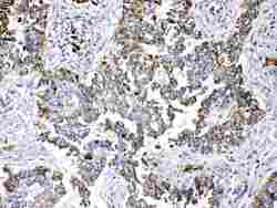
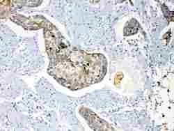
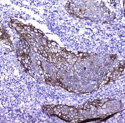
- Immunohistochemistry analysis of Periplakin on paraffin-embedded human lung cancer tissue. Sample was incubated with Periplakin polyclonal antibody (Product# PA5-79856) with a dilution of 1 µg/mL, and developed by Streptavidin-Biotin-Complex (SABC) method.
- Submitted by
- Invitrogen Antibodies (provider)
- Main image
- Experimental details
- Immunohistochemistry analysis of Periplakin on paraffin-embedded human esophagus squama cancer tissue. Sample was incubated with Periplakin polyclonal antibody (Product# PA5-79856) with a dilution of 1 µg/mL, and developed by Streptavidin-Biotin-Complex (SABC) method.
- Submitted by
- Invitrogen Antibodies (provider)
- Main image
- Experimental details
- Immunohistochemistry analysis of Periplakin on paraffin-embedded human tonsil tissue. Sample was incubated with Periplakin polyclonal antibody (Product# PA5-79856) with a dilution of 1 µg/mL, and developed by Streptavidin-Biotin-Complex (SABC) method.
- Submitted by
- Invitrogen Antibodies (provider)
- Main image

- Experimental details
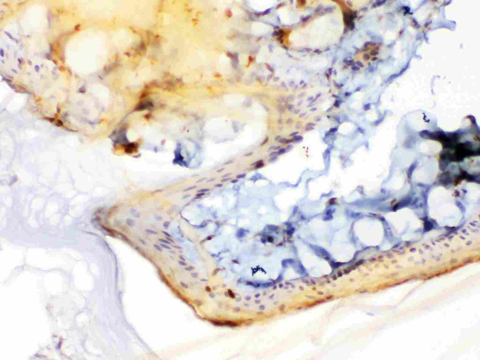
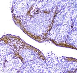
- Immunohistochemistry analysis of Periplakin on paraffin-embedded rat skin tissue. Sample was incubated with Periplakin polyclonal antibody (Product# PA5-79856) with a dilution of 1 µg/mL, and developed by Streptavidin-Biotin-Complex (SABC) method.
- Submitted by
- Invitrogen Antibodies (provider)
- Main image
- Experimental details
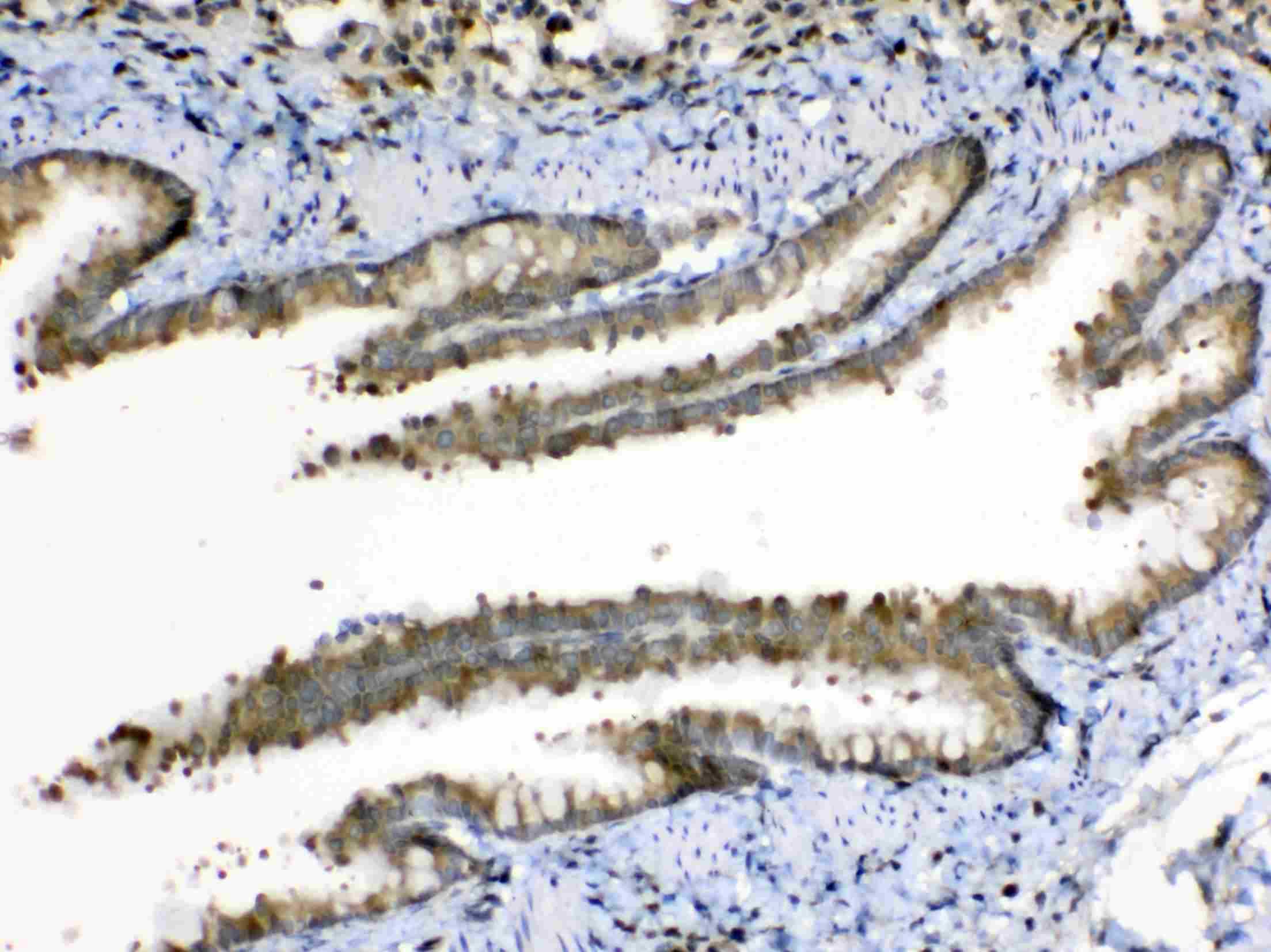
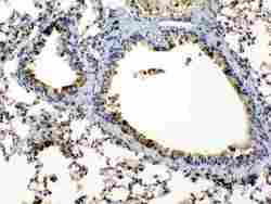
- Immunohistochemistry analysis of Periplakin on paraffin-embedded rat lung tissue. Sample was incubated with Periplakin polyclonal antibody (Product# PA5-79856) with a dilution of 1 µg/mL, and developed by Streptavidin-Biotin-Complex (SABC) method.
- Submitted by
- Invitrogen Antibodies (provider)
- Main image
- Experimental details
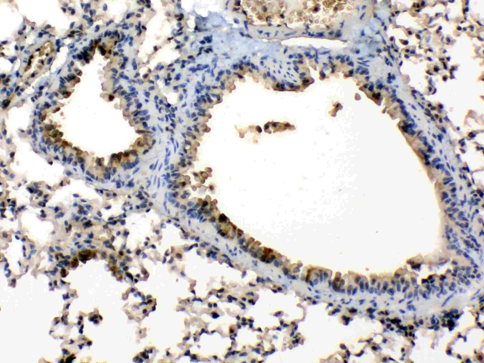
- Immunohistochemistry analysis of Periplakin on paraffin-embedded mouse lung tissue. Sample was incubated with Periplakin polyclonal antibody (Product# PA5-79856) with a dilution of 1 µg/mL, and developed by Streptavidin-Biotin-Complex (SABC) method.
- Submitted by
- Invitrogen Antibodies (provider)
- Main image
- Experimental details
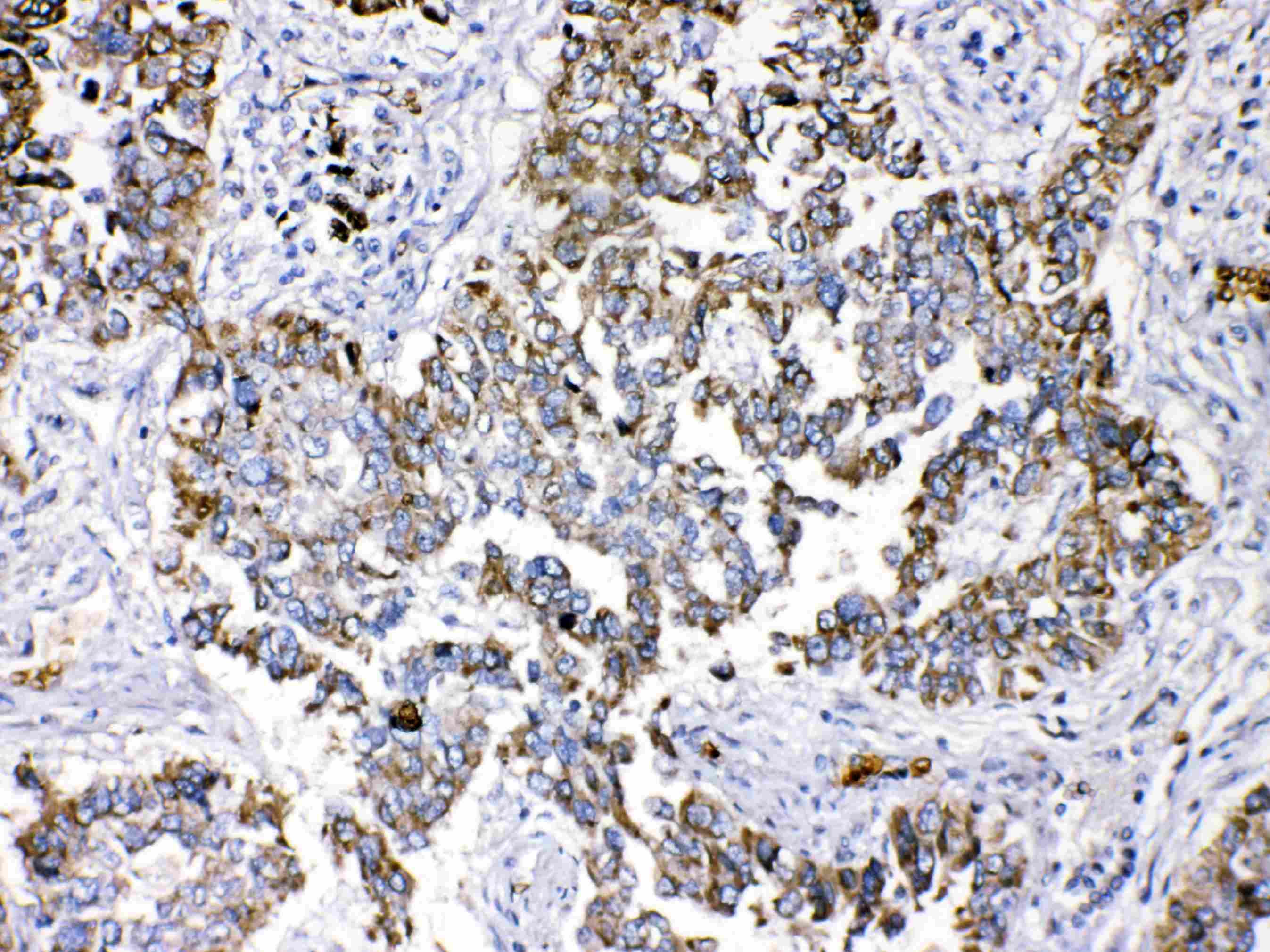
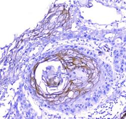
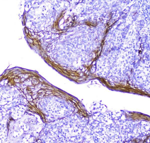
- Immunohistochemical analysis of Periplakin in a paraffin-embedded section of human lung cancer tissue. Heat mediated antigen retrieval was performed in EDTA buffer (pH 8.0, epitope retrieval solution).The tissue section was blocked with 10% goat serum. The tissue section was then incubated with 2 μg/mL rabbit anti-Periplakin antibody (Product # PA5-79856) overnight at 4°C. Biotinylated goat anti-rabbit IgG was used as secondary antibody and incubated for 30 minutes at 37°C. The tissue section was developed using Strepavidin-Biotin-Complex (SABC) with DAB as the chromogen.

- Submitted by
- Invitrogen Antibodies (provider)
- Main image
- Experimental details
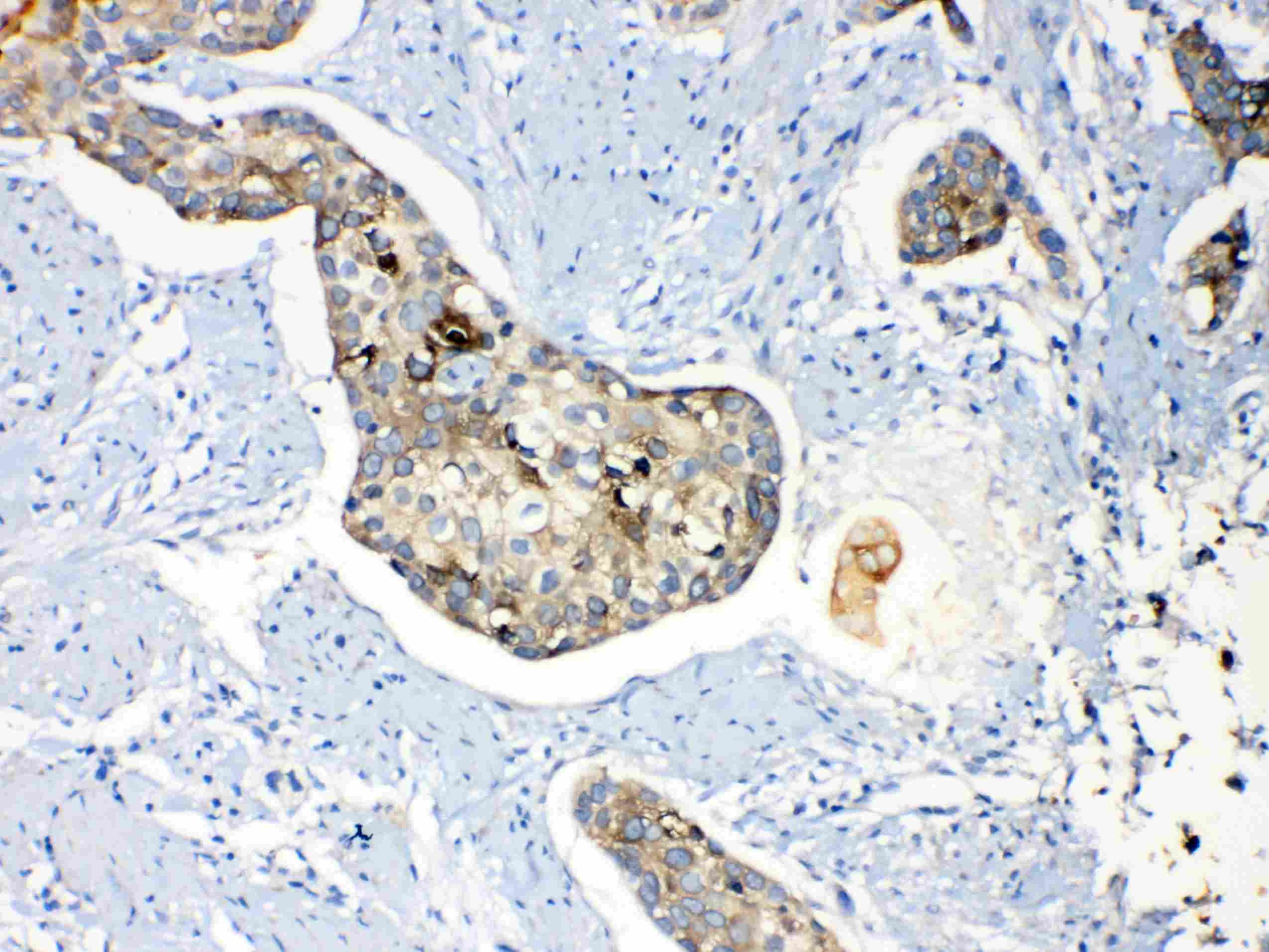
- Immunohistochemical analysis of Periplakin in a paraffin-embedded section of human oesophagus squama cancer tissue. Heat mediated antigen retrieval was performed in EDTA buffer (pH 8.0, epitope retrieval solution).The tissue section was blocked with 10% goat serum. The tissue section was then incubated with 2 μg/mL rabbit anti-Periplakin antibody (Product # PA5-79856) overnight at 4°C. Biotinylated goat anti-rabbit IgG was used as secondary antibody and incubated for 30 minutes at 37°C. The tissue section was developed using Strepavidin-Biotin-Complex (SABC) with DAB as the chromogen.

- Submitted by
- Invitrogen Antibodies (provider)
- Main image
- Experimental details
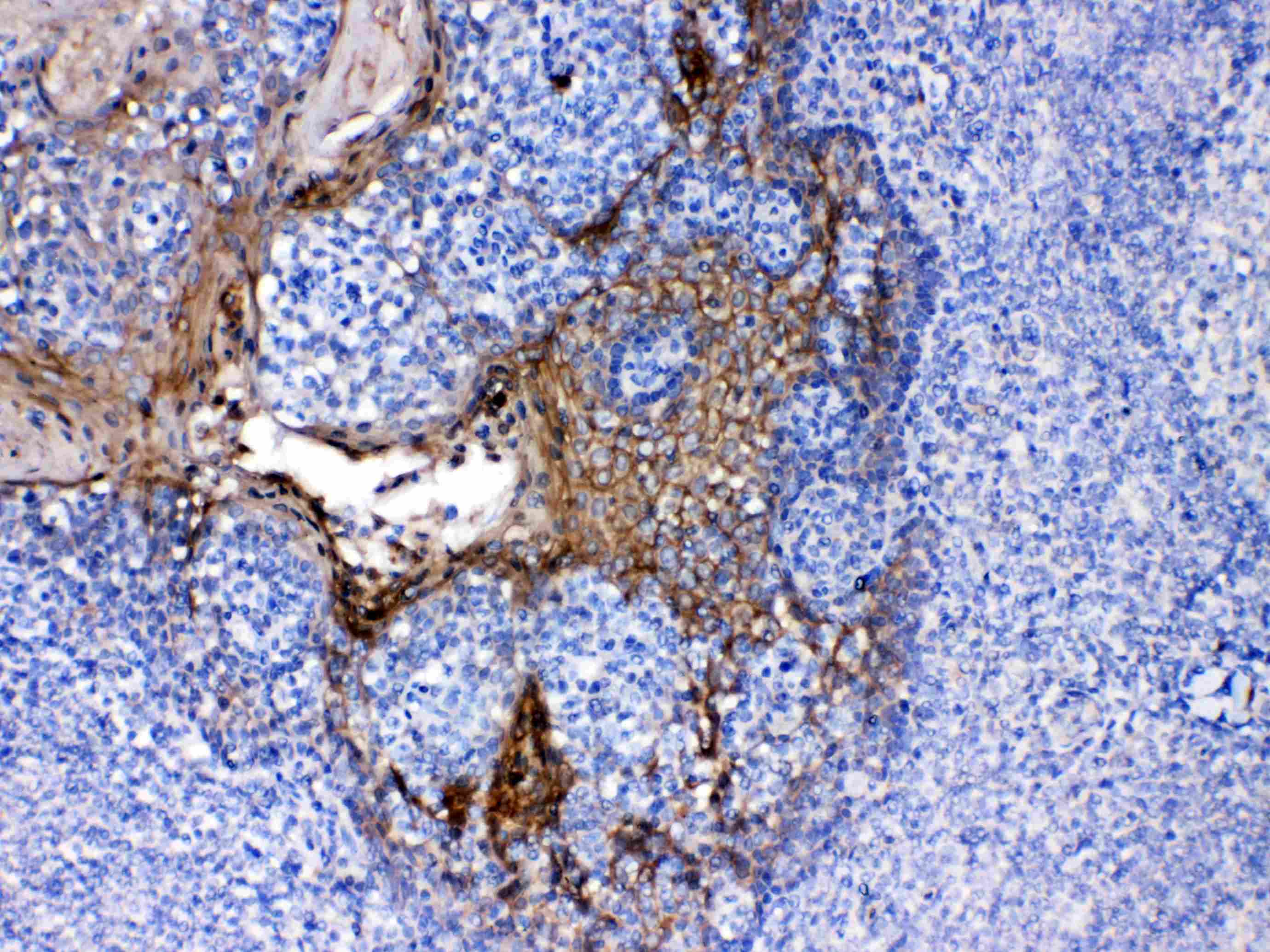
- Immunohistochemical analysis of Periplakin in a paraffin-embedded section of human tonsil tissue. Heat mediated antigen retrieval was performed in EDTA buffer (pH 8.0, epitope retrieval solution).The tissue section was blocked with 10% goat serum. The tissue section was then incubated with 2 μg/mL rabbit anti-Periplakin antibody (Product # PA5-79856) overnight at 4°C. Biotinylated goat anti-rabbit IgG was used as secondary antibody and incubated for 30 minutes at 37°C. The tissue section was developed using Strepavidin-Biotin-Complex (SABC) with DAB as the chromogen.
Supportive validation
- Submitted by
- Invitrogen Antibodies (provider)
- Main image
- Experimental details
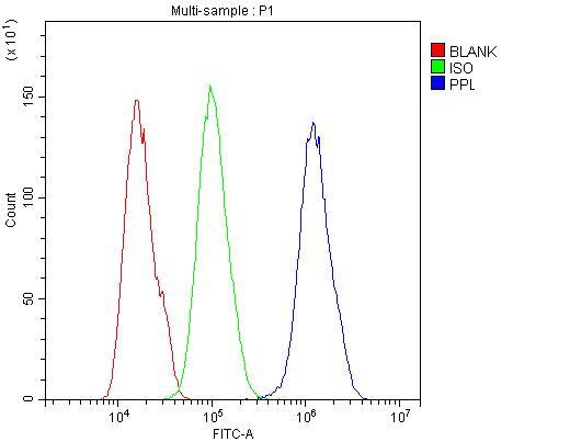
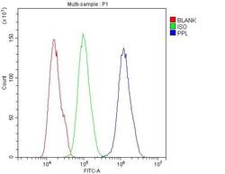
- Flow Cytometry of Periplakin in U2OS cells (blue line), isotype control rabbit IgG (green line) and unlabeled (red line). Samples were blocked with 10% goat serum, incubated with Periplakin Polyclonal Antibody (Product # PA5-79856) at a dilution of 1 μg (per 1x10^6 cells), followed by DyLight®488 conjugated goat anti-rabbit IgG (for 30 minutes at 20°C) using 5-10 μg (per 1x10^6 cells) dilution.