Explore
 Validate
 Learn
Learn Western blot
 Immunoprecipitation
Antibody data
- Antibody Data
- Antigen structure
- References [0]
- Comments [0]
- Validations
- Western blot [1]
- Immunocytochemistry [3]
- Immunohistochemistry [3]
Submit
Validation data
Reference
Comment
Report error
- Product number
- GTX23443 - Provider product page

- Provider
- GeneTex
- Proper citation
- GeneTex Cat#GTX23443, RRID:AB_385050
- Product name
- PER1 antibody
- Antibody type
- Polyclonal
- Reactivity
- Human, Mouse, Rat, Drosophila, Hamster
- Host
- Rabbit
- Storage
- -20°C, Avoid Freeze/Thaw Cycles.
No comments: Submit comment
Supportive validation
- Submitted by
- GeneTex (provider)
- Main image
- Experimental details
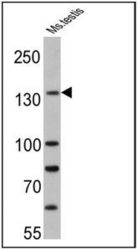
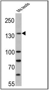
- WB analysis of mouse testis lysate (25 μg) using PER1 antibody at a dilution of 1:500.
Supportive validation
- Submitted by
- GeneTex (provider)
- Main image
- Experimental details
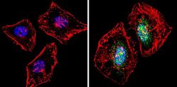
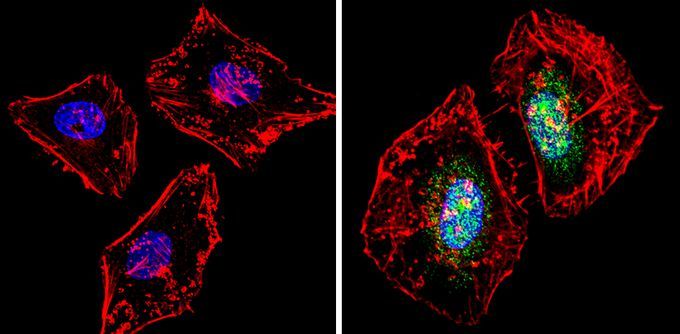
- ICC/IF analysis of HeLa cells was performed in the presence (right) or absence (left) of PER1 antibody at a dilution of 1:100 (green). F-actin (red) was stained with a flourescent red phalloidin and nuclei (blue) were stained with Hoechst or DAPI. Images were taken at a magnification of 60x.
- Submitted by
- GeneTex (provider)
- Main image

- Experimental details
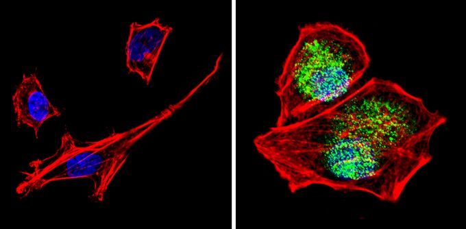
- ICC/IF analysis of SH-SY5Y cells was performed in the presence (right) or absence (left) of PER1 antibody at a dilution of 1:100 (green). F-actin (red) was stained with a flourescent red phalloidin and nuclei (blue) were stained with Hoechst or DAPI. Images were taken at a magnification of 60x.
- Submitted by
- GeneTex (provider)
- Main image
- Experimental details
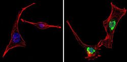
- ICC/IF analysis of NIH-3T3 cells was performed in the presence (right) or absence (left) of PER1 antibody at a dilution of 1:100 (green). F-actin (red) was stained with a flourescent red phalloidin and nuclei (blue) were stained with Hoechst or DAPI. Images were taken at a magnification of 60x.

Supportive validation
- Submitted by
- GeneTex (provider)
- Main image
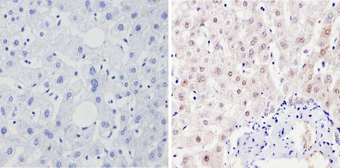
- Experimental details
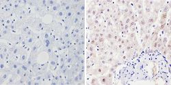
- IHC-P analysis of human liver tissue was performed in the presence (right) or absence (left) of PER1 antibody at a dilution of 1:100.
- Submitted by
- GeneTex (provider)
- Main image
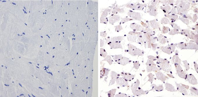
- Experimental details
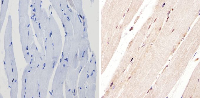
- IHC-P analysis of human skeletal muscle was performed in the presence (right) or absence (left) of PER1 antibody at a dilution of 1:100.
- Submitted by
- GeneTex (provider)
- Main image
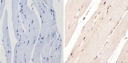
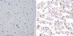
- Experimental details
- IHC-P analysis of mouse skeletal muscle was performed in the presence (right) or absence (left) of PER1 antibody at a dilution of 1:100.